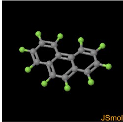
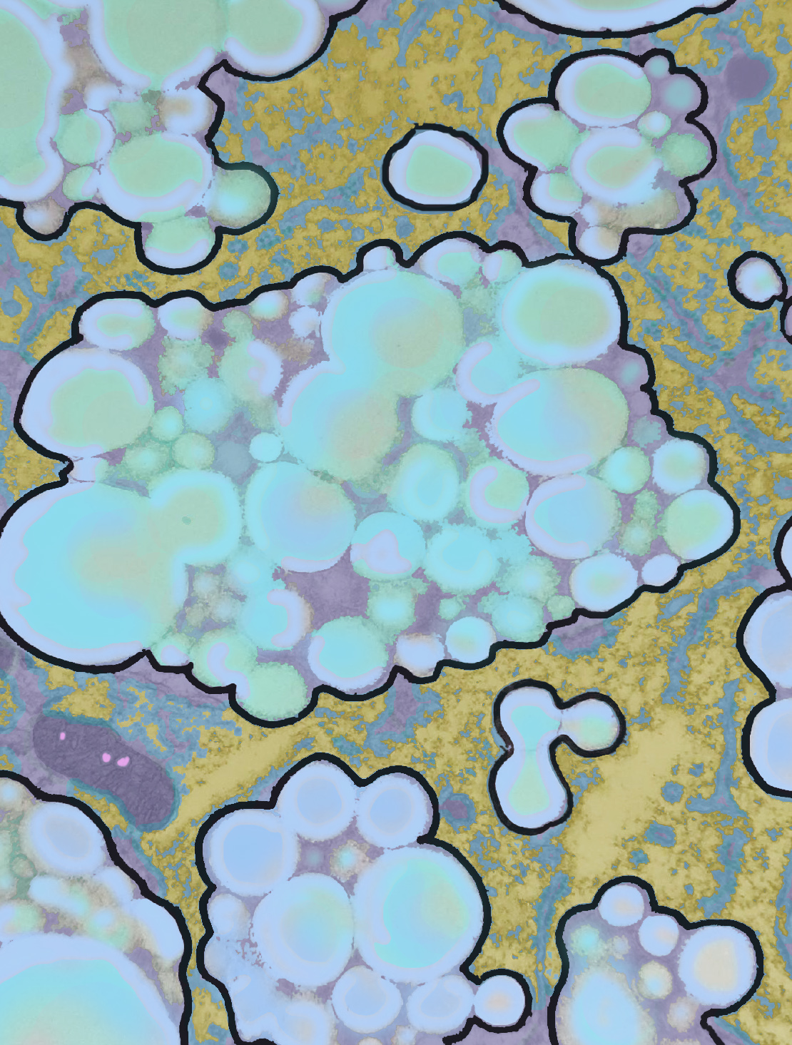
This is a negative I retrieved from storage (40 years past) which has a clear picture of the repartition of perfluorochemical emulsions (just some, dependent on the properties of each perfluorocarbon) within the lysososomes/perosisosomes of liver hepatocytes and interstitial cells. This image is one I pseudocolored showing how within a membrane bound organelle, proteins coat small globules of perfluorophenanthrene (PFP – chemical image from ChemSpider, lined to name).
Image below neg 8946, plastic block 28373, mouse # 26, liver, 21 days after infusion with perfluorophenanthrene (i can look up the dose in mg/kg). This type of intra-organellar, reemulsification of perfluorocarbon based blood substitute is very dependent on which perfluorochemical is infused). Please note that I have drawn around each lysosome/peroxisosome with sharpie (LOL) to define them.
Two mitochondria are present, many polysomes, and rough ER as well. THe pink areas within mitochondria are “not identified as routine cristi, but something has changed there”.
Also note that during processing it is most likely that the actual PFP is no longer there and thus a “ghost” vacuole is present but clearly demarcated by the presence of the proteins fixed within.
