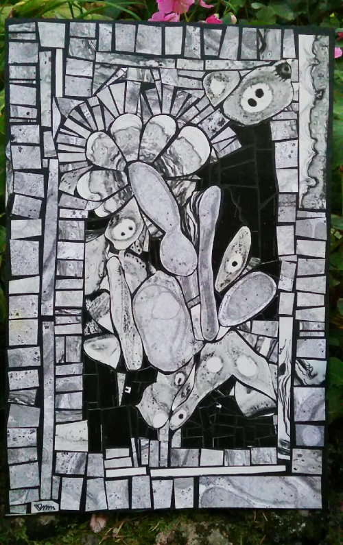
I have used this technique before, maybe decades (more than 3) ago when faced with the very real necessity of pitching my beloved black and white prints from the microscopes I have used in the past, that is light microscopes, electron microscopes, and dissecting microscopes. The tesserae in this picture (which was made expressly for one of my two grandsons) are made from a combination of TEM and LM micrographs. The big-“eyed” structures are keratinocyte nuclei with one or two nucleoli, and one in particular has an apoptotic keratinocyte nearby. There is mouse back skin, and thin layers of keratin present, and some wacked-out cristae formation in mitochondria from liver of genetically engineered mice, and/or rats exposed to toxins for the sake of determining whether we are poisoning ourselves with chemicals and stuff, and whatever else I have saved in the box of potential cut-ups. One mitochondrion in the center has peripheral parallel cristae and the one below that has a collection of protein in the intracristae space. If you look closely you can see the little pale KODAK letters which were on the sides of prints that we used to make of film strips…. that was indeed a long time ago. I actually have saved enough of the extra prints to make a large abstract…. maybe someday I will get to it. In the meantime, identify all the structures that you can.
