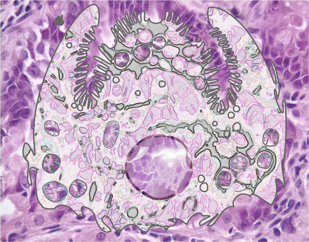
More images of gastric epithelium in the mouse, onto which is layered some vector graphics of mitochondria, vesicles, nucleus (pale condensed chromatin areas within, nuclear membrane (inner and outer membrane and perinuclear space — but no nuclear pores are identified here specifically but the places where outer and inner nuclear membrane come together nuclear pores would be found. This is color and information together, for the 21 century and the digital age.
The microvillar (highly microvillar) apical membrane is shown, the lateral membrane and basal membrane are not distinguished from each other except as the bottom of the diagram is basal.