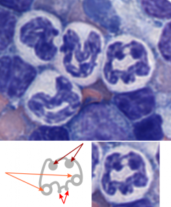
It has been my opinion (based on looking at thousands and thousands of slides and micrographs) that the when the cell undergoes apoptosis or is overly stressed, it shows an amazing nuclear bilateral symmetry. DIVIDED IN HALF (how does this surprise anyone-yet why can I not find this in the literature)– that is to say that that domains in the nucleus which are beginning to assemble, and/or disassemble in response to the cell death signals, such as the interchromatin granule clusters, and the huge nucleolar structures (fibrillar centers and dense fibrillar components) just seem to appear in light micrographs (where sections are relatively thick compared to those for TEM) in a mirrored symmetry.
I have seen this same phenomenon in white blood cells (particularly bone marrow smears) where the granulocytes were stressed and micronuclei, or bar-like bodies, or segmentation are increased by (in some cases in bone marrow smears from Cyp1A1, Cyp1B1 and Cyp1A2 ko mice and their many variations and exposure toxins like benzo(a)pyrene or TCDD), and in other cases such (in vitro) where RNAi knockdown of the highly conserved anti-apoptotic gene, C9orf82, induced apoptosis and cells showed mirrored nuclear symmetry, and in 14CoS cells that underwent massive synchronized hepatocyte apoptosis.
This is part of the symmetry of the nucleus currently being explored with data mining, fluorescent antibody imaging, TEM-CT, and yes, even the old school “eyeball” approach. Here is an image and diagram of one bone marrow smear where bilateral symmetry of a white cell is very obvious, and though the image is almost a decade old, and I queried the literature at the time I could find no publications on domains, territories, or mirrored images in apoptotic cells. (BTW the cells below in a bone marrow prep are NOT apoptotic but nicely segmented).
