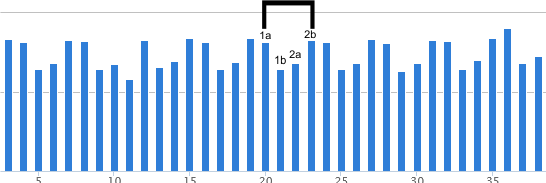
Does anyone see a pattern here, in the glycosylation peak height, arm 1 and arm 2. It is very clear that the different image analysis filters dont mean a whole lot, when compared to the actual molecule, the relative heights of each of the peaks along a length of a trimer (hexamer = 2 trimers). This made me examine the trimer glycosylation peaks of this SP-D dodecamer separately, comparing peak heights. Repeating pattern for each arm is clear. Since each hexamer is plotted as trimer 1a and 1b, and 2a and 2b, the fact that the difference in the hexamer glycosylation peak heights is not significantly different using a t-test, even though this pattern occurs, I am going to continue to use the data each trimeric arm separately.
It is important for me to say that I think thee are differences in the glycosylation peak height that relate to the “number” of arms in the trimer that actually have a glycan attached — could it vary, one, two or three, which would then result in a different grayscale value for peak height (and valley).
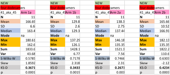
 The mean for each glycosylation peak in each of the four trimers of the dodecamer, and each of the image and signal processing apps (n was 11 for each trimer)
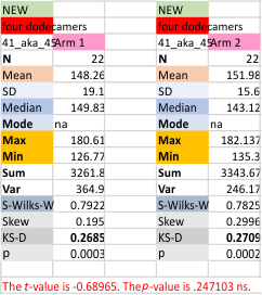
 t-test comparing the four trimers follow.

Values for the glycosylation peak grayscale height for each hexamer of the single dodecamer (that is arm 1a and 1b, and arm2a and 2b) in a t-test were not significantly different).
