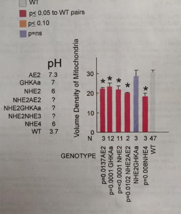
These data are all performed on mice (KOs and treated) from the laboratory that was run by Dr. Gary Shull and his students and postdocs. I saved these plots (which were intended to be submitted as a manuscript back in 2007, when I retired) and am just now sorting through the data from those years in microscopy. I did morphometric measurements on many of the mice (listed below) which were published, but gathered together those into a composit kind of analysis — not a publication, just a view into what could have been published had time permitted. I dont think at this point anyone will try to rebuild this, but the information as a “glimpse” into changes in the basolateral (i dont like that term, it is clearly a lumping together of two very distinct membrane domains, nevertheless it is in common usage), apical membranes, tubulovesicular, and other structures into a quantitative assessment over many different genetically modified, or treated mouse parietal cell datasets.
The graphs in the pdf represent hundreds and hundreds of hours just for the morphonetry, let alone the animal experiments, the creation of the animals.
The plots in this pdf (some shown below just so you know what is in the pdf) are low quality prints that I used just for reference. The actual data can be recovered with great effort but if someone wants the numbers, i will try to retrieve the original datasets. A pdf of all the graphs is HERE parietal_cell_morphometry_many_KO-mice1
Selected screen prints below”


