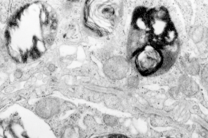
Electron micrograph of an alveolar type II cell from a lung lesion in a guinea pig M8035 ( a study in the mid 1980s and vinyl chloride inhalation and extra vitamin C) showing a banded protein structure which i have to assume is a collagen, since the section provides a continuous tracing to the microvillar surface of the cell. What looks to be an intracellular object does NOT fit the parameters for an intracisternal body since the ribosomes are not at the growing end of the object and it can be traced to alveolar space, even on both ends. But it is also not typically what looks like basement membrane…. So YOU can name it, I will think about it.
