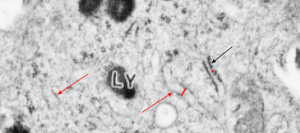
CUTZ, WERT, NOGEE, MOORE. Deficiency of Lamellar Bodies in Alveolar Type II Cells Associated with Fatal Respiratory Disease in a Full-term Infant. American Journal of Respiratory and Critical Care Medicine, 2000, Vol.161: 608-614 paper shows two electron micrographs which examined as closely as the images would all at their printed resolution. The specifics I was looking for surrounded two different styles (types, configurations?) of RER. OF course the hope was to find RER with some kind of layered protein inclusion granule that might suggest that a surfactant protein was being overproduced, but nothing like that was found. It would have been reasonable I think, to expect that if any surfactant protein was being overproduced there would have been RER profiles. The RER profiles in their higher magnification electron micrograph did show more RER (and some dilated rounded RER) than a typical alveolar type II cell has, and as they point out, more, and more varied types of MBV and lysosomal? endosomes. I was hoping to find, in particular, RER profiles expanded in a long dimension with a width of about 100 nm. Their magnification marker was not applicable to the published pdf so I have marked in red a single ribosome and assumed it was between 25-30 nm in diameter. THen placed these perpendicular to the wide-dimension of two adjacent RER profiles. One you see is a typically closely applied ribosome studded profile, the other is approximately 100 nm in width. I wasnt able to conclusively see any central band within that wider RER profile, let alone 3 or 5 or 7 layers. Here is their pictures (used without permission..but it is open source). They marked the MVB/lysosome (Ly) I marked the single ribosome (tiny red dot) and the span of 5 red dots across the wider profile of RER, and the two red arrows to wide RER profiles and the single black arrow points to a more typical less wide RER profile.
