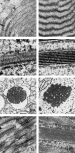
I was looking in the literature for examples of layering or high organization of proteins within the RER of various cells. There is no dearth of examples. Many really wonderful photographs are found in the book edited by Feroze N. Ghadially. Ultrastructural Pathology of the Cell and Matrix: A text and atlas of physiological and pathological alterations in the fine structure of cellular and extracellular components, Edition 3, Volume 1, Butterworths, London, 1988 THESE IMAGES ARE FROM THAT BOOK AS AVAILABLE ONLINE, JUST SCREEN PRINTS AND CROPS (without permission btw).
 There are some very awesome pictures which show enormous variation in the orderly arrangement of proteins (mostly in pathological situations, as is likely for the alveolar type II cell that might be packed with surfactant protein A in the RER). Some show some of the characteristics of become ordered at the transition from RER to golgi, sort of signifying that something has be modified in the protein, or the RER microenvironment that has allowed for alignment of the protein molecules. A, banding with heavy lines and very precise spacing B, bands or layers of very different densities – and also widths C, tubular appearance C, D, central aggregagion spacing after protein synthesis, abundant ribosomes on the whole granule (unlike the alveolar type II cell granules where there is a definite directionality) E, F, parallel and perpendicular directions G, and numerous layers H.