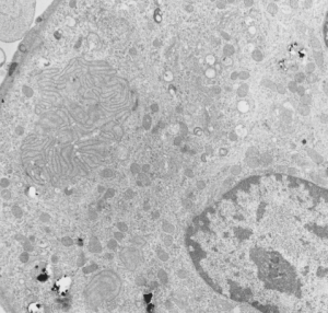
Rabbit lung: post intratracheal instillation of iron oxide and dibenzo cabazole, electron micrograph. This is not a great picture, but i post it just so one can appreciate that there an area of odd membrane swirling in the middle left, and a small area just like it in lower left. The cell has a lobular nucleus and there are iron oxide (probably with DBA alongside) within this cell. I don’t think it is an alveolar parenchymal cell, but likely an inflammatory cell. A type II cells from the same microgrpah has lamellar bodies, which this cell does not, which further indicates it as a migratory cell. Animals given DBC do get tumors. neg 4447, block 17793, Rabbit #32-3. Sorry the micrograph has stain ppt on it, but it is worth looking at just for the layered structure.
