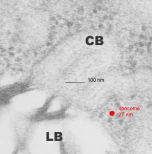
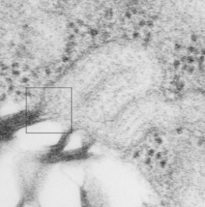
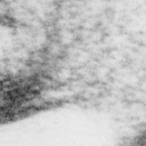
Margin of a cisternal body (RER granule) and a lamellar body in an alveolar type II cell gets blurred in this electron micrograph. There is a transition area right at the point of blending (LB, lamellar body; CB, cisternal body (or granule with what I hope is surfactant protein A all oligomerized in a long protein extruded order). The view at the transition does contain a lot of hexagonal structures, which have a reasonably good fit for the 26 nm given to the bouquet top of surfactant protein A and a nearby measured ribosome (mammalian ribosome about 27 nm)… there is some kind of linear alignment too, right at the interface between the two organelles…. any speculations. Top figure (unretouched) shows ribosome for size comparison and a 100 nm bar marker, a granule with three periods pointing from the lamellar body to the upper right corner of the micrograph. The linear interface lines are interesting, but one can see the ribosomes (burned in photoshop in figure 2 (middle figure) as well as a burning in the hexagonal pattern which occurrs just ad the interface of the lamellar body and the granule. Box in middle figure denotes inset as bottom figure, where the hexagonal pattern is more clearly seen (as I enhanced this with the burn tool in photoshop. 9856_17085_guinea_pig_#301.


