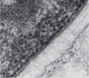
Typically the outer nuclear membrane of mammalian cells has ribosomes attached to it, the inner nuclear membrane has lamin, which appears as a band on that inside membrane. The two sides of the nuclear membrane connect at nuclear pore complexes (not seen in this tiny portion of the nuclear inner and outer nuclear membranes of a guinea pig type II cell (separated by a light band of intracisternal highly organized protein which also can fill cisternae of the rough endoplasmic reticulum in this species under some circumstances) – see other posts in February 2016 regarding that protein.
The inner nuclear membrane, its integral membrane proteins, plus the lamins (the latter lacing the nuclear pore complexes together) participate in DNA replication and RNA processing along the heterochromain – euchromatin interface.
While working on describing the protein contents of the space between inner and outer nuclear membranes I ran onto this really wonderful electron micrograph of a tiny portion of an of the edge of a nucleus where there were very apparent “loops” — in the nuclear structure — maybe indicative of some sort of DNA or RNA activity (looking distinctly like beads on a string). This is a configuration which matches several diagrams found online for active (green) and inactive (orange) sites for DNA synthesis (or maybe RNA processing?) within the nucleus. I couldn’t resist trying to highlight this structure in color — at least as I saw it.
