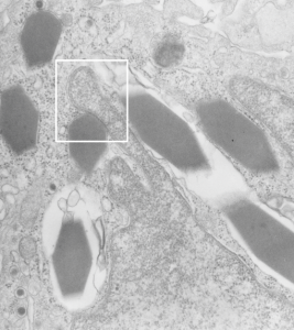
Looking at an alveolar macrophage from an aged guinea pig without treatment (a control) I noticed a cell which contained some very nice crystals, but noticed in particular that when a particular crystal was juxtaposed to the nucleus, that some kind of “order” appeared, and an increase in density of the nuclear membrane was evident. This would be an interesting thing to research if the macrophages with these inclusions were frequent. At best, I have seen only a few. As usual, negative 9218_block17082_gpig_301_lung, likely alveolar macrophage, data given just in case someone out there wants backup.
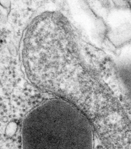
White box is enlarged and contrast enhanced below this image. Nucleus is a finger like projection from the lower right hand side, and at least 7 crystals are seen.

 I can visualize these little A-frame things with heads on them about 10 of them on the inner nuclear membrane. Outer nuclear membrane and granule membrane is really indistinct… owing to the ribosomes in the limiting membrane of the crystal, one could even assume that this crystal might be in the perinuclear space.