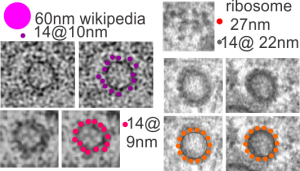
Found these two pretty nicely organized COPI I (probably) vesicles in an alveolar macrophage from a mouse which breathed E2 bubble oxygenated liquid for 3 hours and was allowed to recover for 48 hours. This macrophage had particles (droplets) of E2 within but I cropped out the two vesicles which displayed pretty even symmetry and a close to equatorial sectioning.
These micrographs were all marked up with pen and lines and I rescanned it from the negative (1347_4840_2,500x, mouse, E2 breathed 3 hr, 48 hr recovery, electron micrograph).
I sought out some TEM images of COPI I vesicles and found a composite of in vitro vesicles on wikipedia here, and from images, this article on researchgate which offered up some info as well. I think my images show the best symmetry, just by chance at about 14 densities per approximate equatorial circumference. Diagrams of COPI I vesicles don’t seem to offer an exact number of constitutive or incidental “blips” on the cytoplasmic side of the membrane, so this is just a guess. My dimensions were derived from adjacent ribosomes, taken at 27nm each as an average, Their calcularions?? but as an estimate perhaps those densities on the surface are 10-14nm in their rounded diameter.
