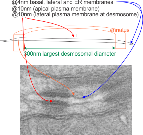
Looking at a summary of desmosomes (so called macula adherens — spots of adhesion) one quote was “randomly arranged” which probably is naive. I find desmosomes (in the only tissue where I have looked seriously… hepatocytes, from several species) to something around a half to a micron, when found cut in the widest diameter. (that diameter There are many instances where the distance between the desmosome and the microvilli of the bile canalicululi is occupied by an adherens junction and a tight junction (pretty much common to epithelia). This is the point where i would have issue with the comment “randomly arranged”, in my experience (which is only limited by my age, occupation and brain) suggests that “very little in biology is random”. (check out this site for bio-numbers) that says the trilaminar membrane is between 4 and 10nm wide.
It is pretty clear that the intercellular space of the desmosome is constant “height” (approx 10nm) as opposed to the dimension across the diameter (approx 300nm at the widest point) which varies depending upon whether the cut is at an equator or tangential to the desmosomal spot.
It is pretty clear that the plasma membrane within the diameter of the desmosomal proteins is also rigid (owing to some kind of stabilization perhaps because of the transmembrane domains of the cadherin proteins that make up the intercellular portion of the desmosome.
It is also pretty clear that whatever is present in the outer mitochondrial membrane (some kind of link and presence of some type of intermediate filaments) that tether the mitochondrion to the plaque proteins of the desmosome also creates a rigid appearance to that membrane.