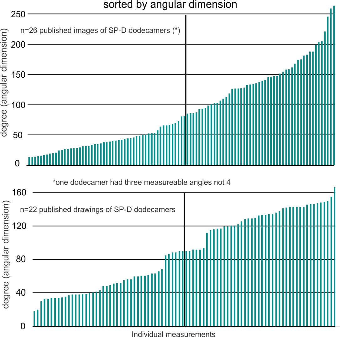
Trying to determine where there is any rhyme or reason for the angles that individuals choose when they diagram SP-D dodecamers I decided to compare some actual measurements vs some drawings. Each and all images come from published (peer reviewed) articles available to anyone who searches the internet for surfactant protein D.
Briefly, there seems to be a huge variation in angular dimension in the photos taken using various types of electron microscopy (including atomic force microscopy), more variability in fact than the diagrams would represent. Initially, the diagrams that represent SP-D as an X with 4 90o angles are not the most accurate, as the “mode” for angles from measuring “real” images is about 30-50o and 120-140o. Because of the variability in angle dimensions (and a publication by Arroyo et al demonstrated something along these lines using pH as a variable to alter the angular dimension of SP-D dodecamers (and multimers)) one has to think about environmental factors in the shape of this protein. In the search for a good pattern recognition protein to attach to nanospheres for immunity… the hunt for the optimal angle(s) is important.
Below is the set of measured angular dimensions for SP-D. A summary sheet for the “real” SP-D images from several different authors (including, Waters et al, Crouch et al, Hildago et al, Hseih et al, Ohya et al, Kishore et al, Arroyo et al, to name a few) follows. It becomes clear that the division of SP-D dodecamer arms into acute and obtuse angles as portrayed by the authors in their diagrams may not replicated in the real life continuum, likely due to environmental influence (including pH as described by Arroyo et al, but other factors as well).