It is clear that when TEM preps are made that not everything lies down on the grid in perfect orientation and without blemish or wrinkles. The mess that comes with looking through the microscope is not going to change much, it behoves us to make use of the visual cortex of the mind while waiting for AI to become more sophisticated at recognizing shapes and grayscale.
So SP-D trimers (from a picture by Arroyo et al mentioned countless times) are not all the same length (some folding over or back or side to side -probably at the N terminus) and not always the same thicknss (shrinkage and or slight perspective in depth) direction of the cantilever in AFM, and they are not all the same spectrum of grayscale (either 0-250, 1-256, or 1-100% your choice).
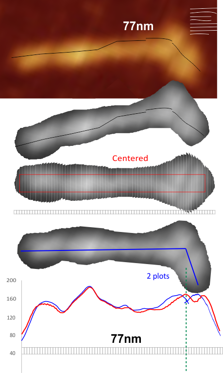
The trimer length measure below by discontinuous lines (to accommodate the bend) and images converted to 300ppi grayscale, and those grayscale images exported to RGB tiff files to import into image J. One image as you can tell has been cut into 77 pieces, which should equate to about 1 nm each. Those slices were centered and the image exported to tiff for LUT plots using ImageJ. (too bad the irregular polygon select tool wont plot in imageJ haha..). The other image was analyzed in 2 plots, one along the straight part of the arm, the other along a straight part of the arm rotated. Two plots were scaled the same and just cut and pasted into the first plot. Red rectangle and red plot line are from the centered image, while blue is from the plot from two areas, aligned at the matching plot indices.
It is pretty clear that both methods produce similar results, the former is perhaps easiest.
Bottom line for this particular SP-D trimer appears to be that Nterminus is a separate peak (albeit about half the height (lightness) of a hexamer or dodecamer) ) which suggests that maybe there is side to side binding (end to end would create TWO PEAKS WHICH ARE CERTAINLY NOT SEEN.