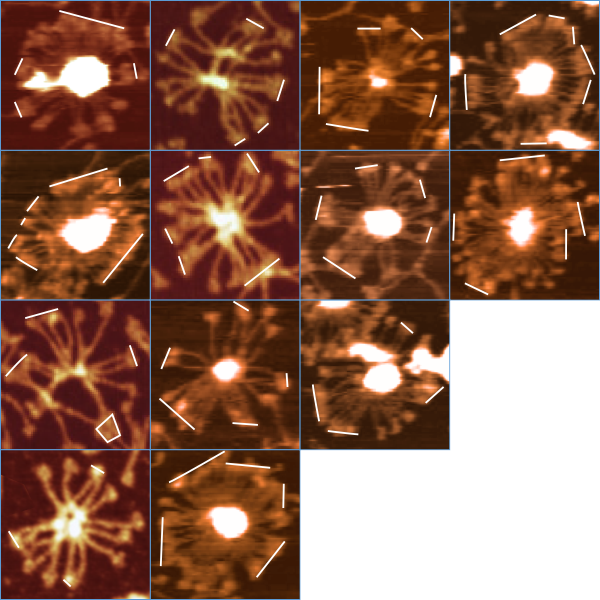
I have wondered about this observation for two years. Why are the CRD in multimers (and also sometimes dodecamers) somtimes arrayed in an obvious “lineup”. Very often the CRD in separate trimeric arms that are adjacent in the outer sphere of fuzzyballs appear to cling together. Also, there are events which (maybe during processing only, i doubt anyone has mentioned it because it could have been a processing effect) where many CRD seem to be aligned and the angles between the aligned arms is obvious.
In all microscopy (and in all other media preparations, bar none) there are admittedly artifacts which appear. Here are images from a couple of authors, separate procedures, but the same obvious event. While it may be accentuated by processing, my guess is there is more to the CRD clinging than one wants to contemplate.
AFM is not my field of microscopy, but there have been few artifacts that i have seen in TEM that dont also provide information about cells and tissues in general. Moreover, in LM, the artifacts are horrific, yet they are given credence daily by a host of pathologists. The sticky CRD are a clue, to what? It is obvious too that the greater the number of trimeric arms, the more apt there are to be groups.
Below find a montage of SP-D images from various authors upon which i have marked the most obvious “line like” associations of CRD. Just for convenience they were standardized to similar diameters (@ 150nm is a good estimate).