No complaining here but sometimes it is hard to see progress in laborious tasks, and maintain forward momentum.
Case in point, trying to find out the number of “height” “brightness” “LUT” “grayscale” peaks in surfactant protein D.
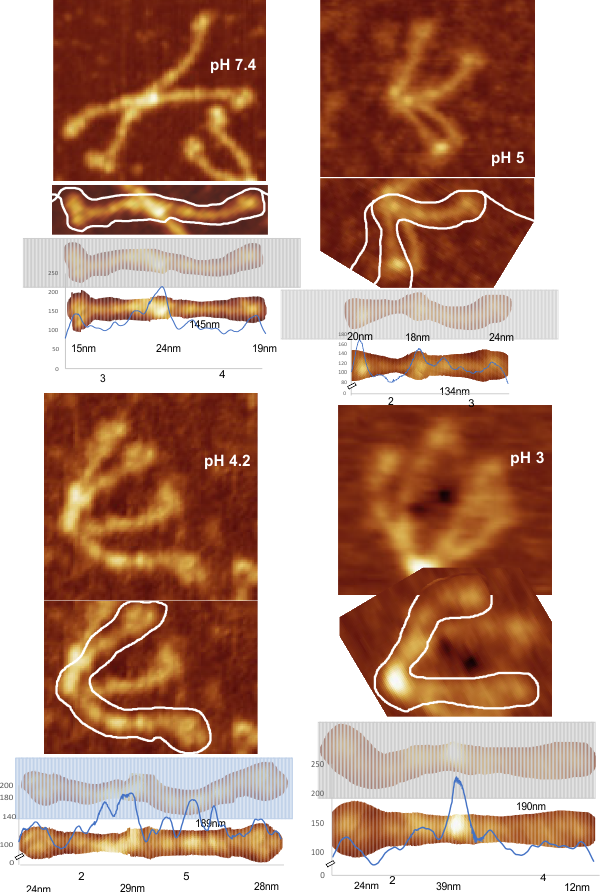
The publication i have quoted the most is by Arroyo et al, and again it is here along with an assessment of four of their images
for peaks along the collagen like domain of the trimers. All the techniques previously employed to create easy to measure, to align, to adjust to a given-consensus-diameter, have been explored before in this blog, but this looks at other parameters. Arroyo et al subjected isolated hSP-D to different pH conditions and as I looked over the images i realized that pH apparently does not affect the presence of LUT peaks along the collagen like arm, thus i isolated, rotated, cut into 1mm slices and centered (horizontally) these images, and then subjected them to FIJI upversion of old ImageJ.
One of the good things about dicing and rotating and using two arms of the dodecamer is that one quickly realizes that there is relatively small effect of bounce, chatter, skipping, vibration in the plots of grayscale. In fact, in all these plots images taken at different angles to the trimeric arms, different magnifications, different resolutions, different HSL, different brightness, and contrast, and at least three different methods of sample preparation, and almost a half dozen different species/genotypes of SP-D really dont change the pattern of the LUT plots that much. That is consoling.
Image below has four images by Arroyo et al, and in each i have plotted a hexamer, and in each i have shown what processes i have used and given the visual peak number and shown the actual peaks created. In the 8 trimeric arms there is a mean of 3 peaks between the N termini and the CRD peaks. I have shown the nm measures for the N and CRD in each of the four samples.