After having looked seriously at many many SP-D images (AFM, rotary shadowed, negative stained) and the shape in LUT plots at the end of each dodecamer so consistently have a teardrop shape that it is hard not to make the association between that and the partial molecular modeling of SP-D.
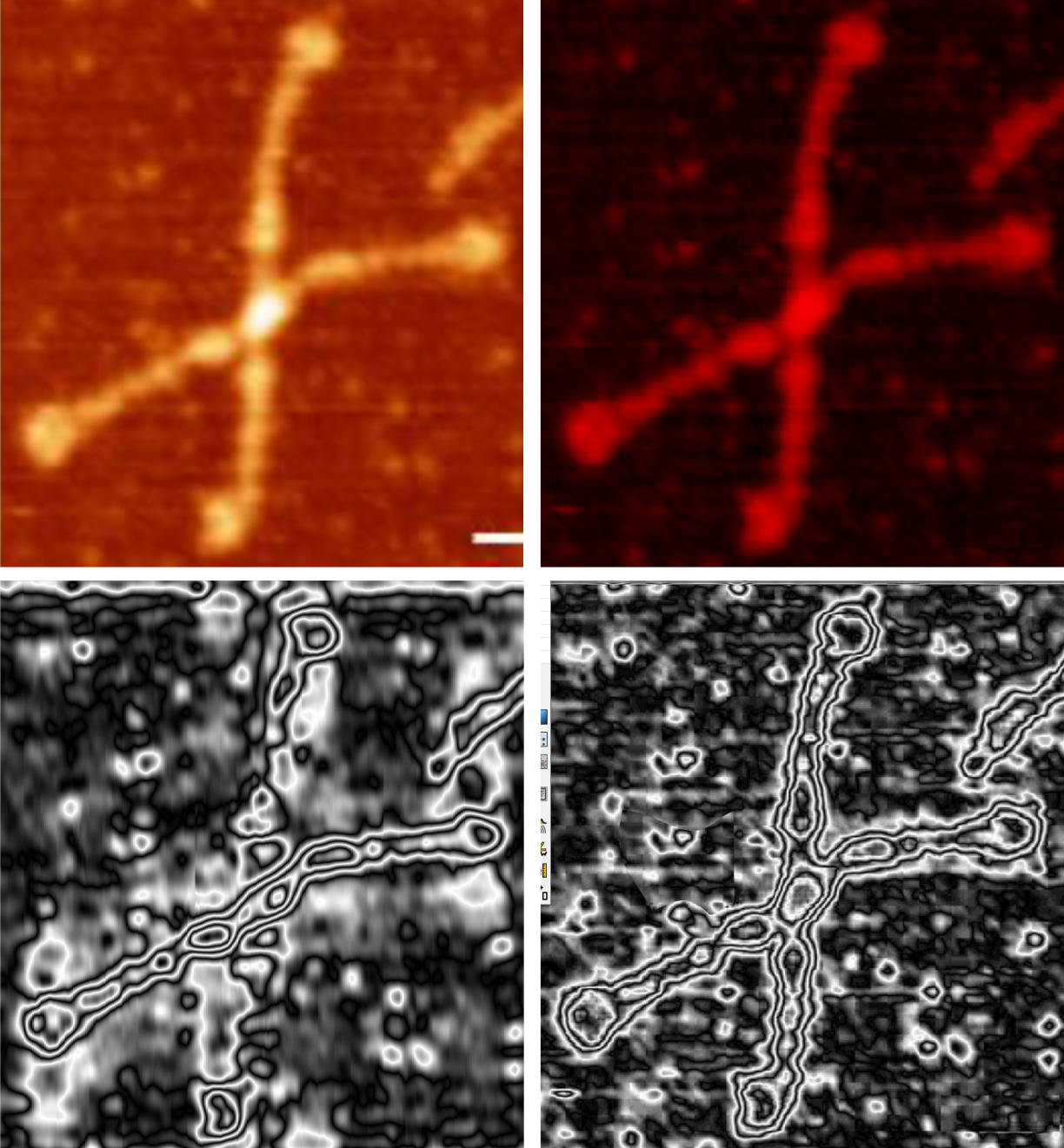
It is really hard not to see the correlation. Top four images are the original from a figure by arroyo et al (top left), RGB saved from gwyddion, bottom left, 1D fft, bottom right 1D horizontal fft (i dont know much about fft (yet), but it is clear that the teardrop shape does not go away with many kinds of image processing. I may represent the folded distance in nm of those two domain (neck and CRD). Another thing that is common is the narrowing of the neck coiled coil at the change to the collagen-like-domain. One can contemplate whether the collagen like domain is more tightly wound than the neck as seen in countless images of DP-D and whether the dimensions from the edge of the CRD to the neck can be measured using plots.

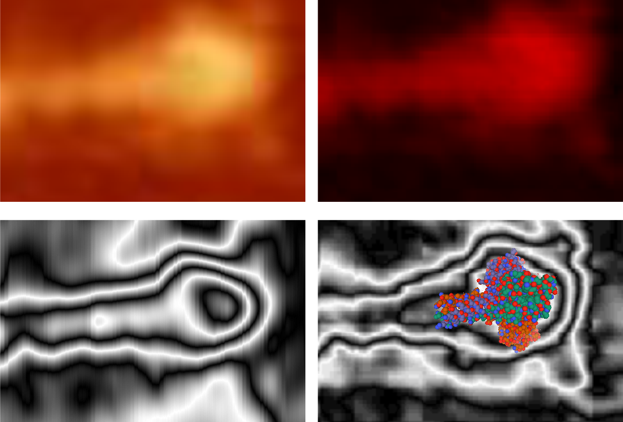
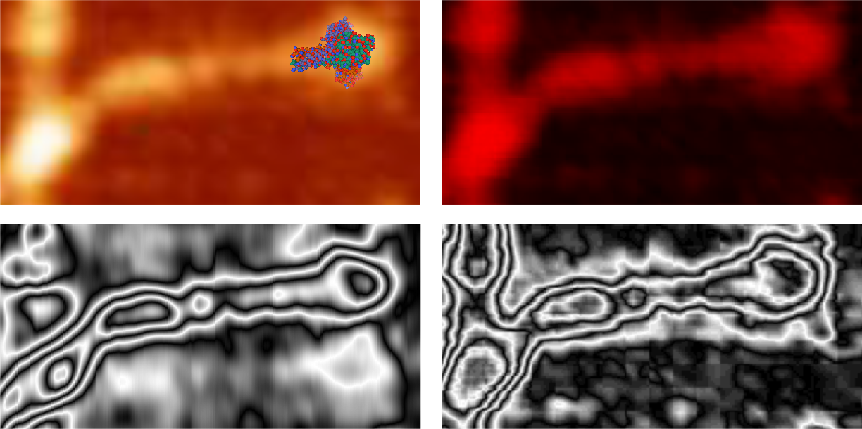
Panel below is an enlargement of identical segments from each of the four pictures above, one has overlay of SP-D neck and CRD, as imaged in RCSB.
Bottom panels are portions of middle panel, just enlarged and with molecular model overlying (in this case) the vertical fft transformed image. 
