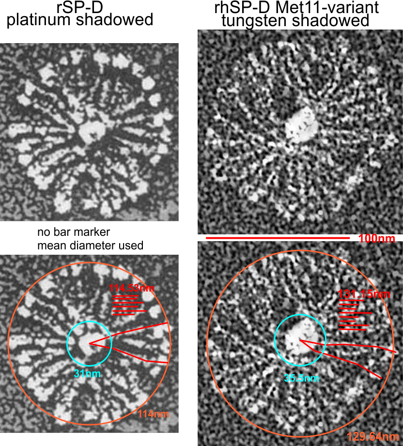
Two shadowed images (top left published rat SP-D, 1994, Ed Crouch et al) SP-D multimer. Methods mention platinum and 10o angle. There is a lot of detail that I particularly like, mainly the dark area around the N termini group and the bright peaks (making a ring at about 35nm from center) and even in many of those arms there are smaller concentric rings that I would hope someday will turn out to be equivalent to the brightness peaks that occur routinely in AFM images of SP-D. Image on the bottom left is my measurement of the top left image. There is no bar marker on the set of images from the publication by Ed Crouch et al but they do give a mean diameter (CRD to CRD) of 114nm. I measured two of the arms to the center point with a segmented line and came up with an almost identical dimension (see lines and nm values on bottom left images).
The top right image is a recent photo sent to me by Jack Griffith, made from recombinant human SP-D prepared by Grith Sorensen. The method of highlighting the multimers (detailed in Ed Crouch’s manuscript – where they used a gray lettreset screen over the entire image then brushed away the gray screen in areas that were just the SP-D arms to highlight the molecule) I basically copied on Jack Griffith’s image however i used Photoshop, and a 45% screen and the eraser tool to highlight the arms of rhSP-D in the image. (Both images are inverted from their originals). There are interesting differences and similarities. Size is one difference, and perhaps the angle of shadowing and the amount of shadowing in the left images gave me the impression that the CRD were a lot bigger (nm in diameter) and a greater part of the arm of each trimer than they apparently are. CONSIDER differences in species/SP-D size…. and protein preparation and shadowing techniques. (left images ratSP-D, right images recombinant human SP-D).