Category Archives: Ultimate order, the cell

The beauty and order of life is astounding.

What part of the collagen-like domain of SP-D forms the center with the N termal?

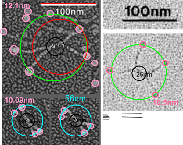
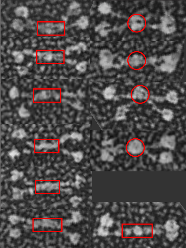
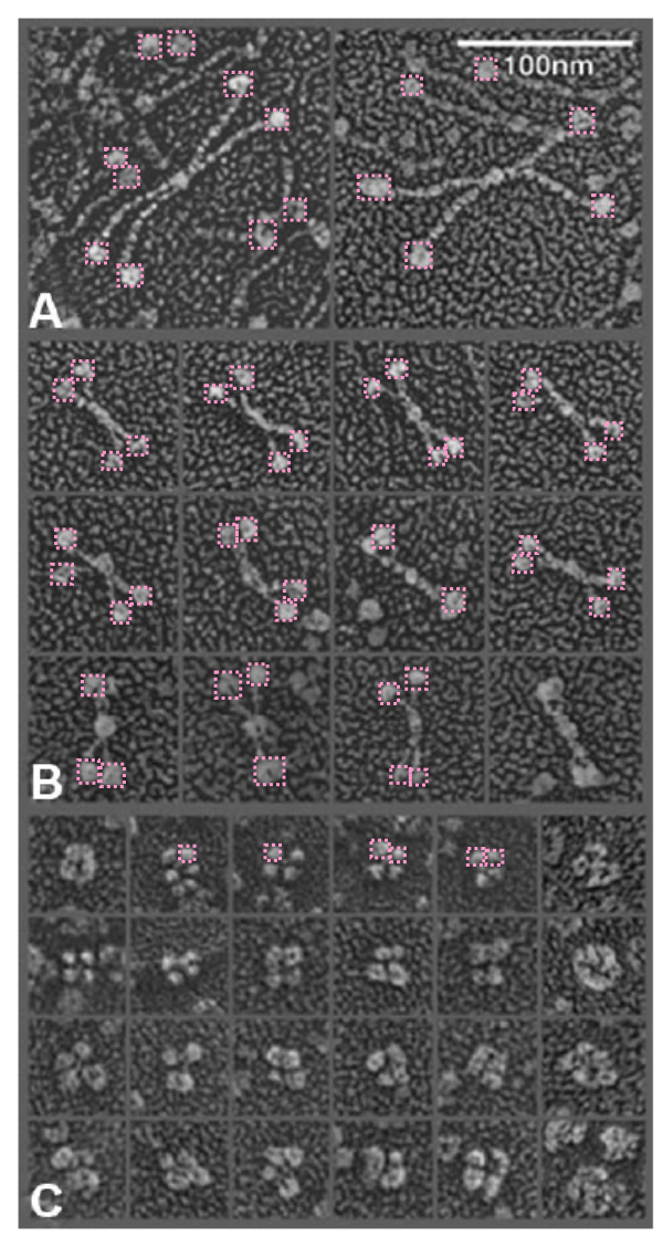
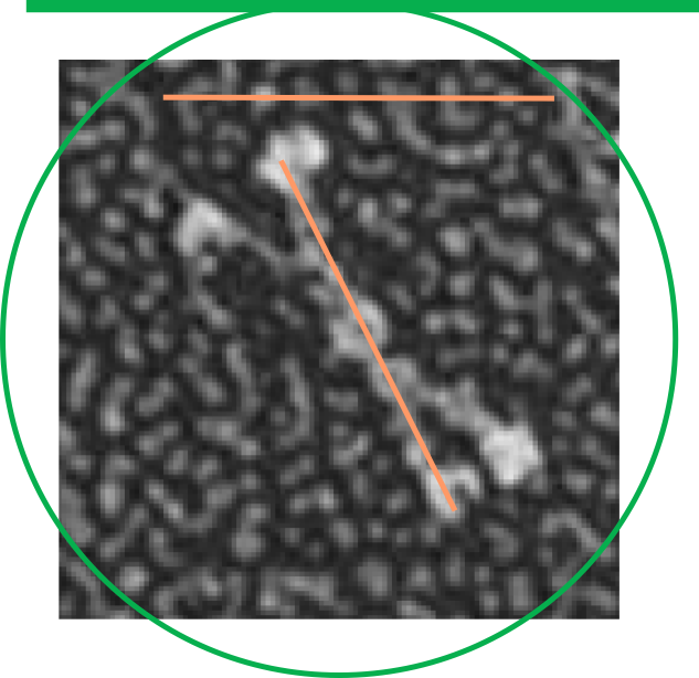
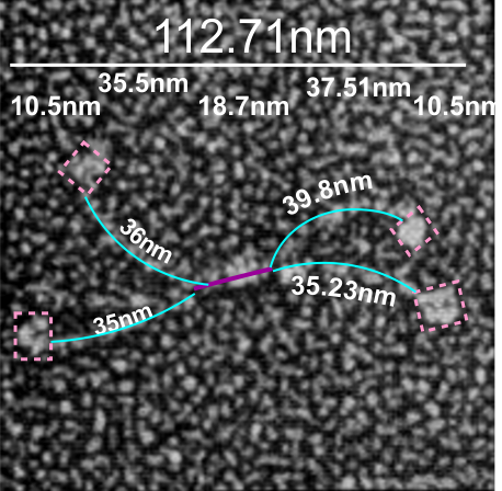
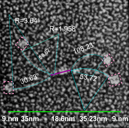
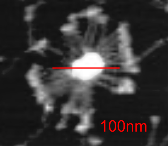
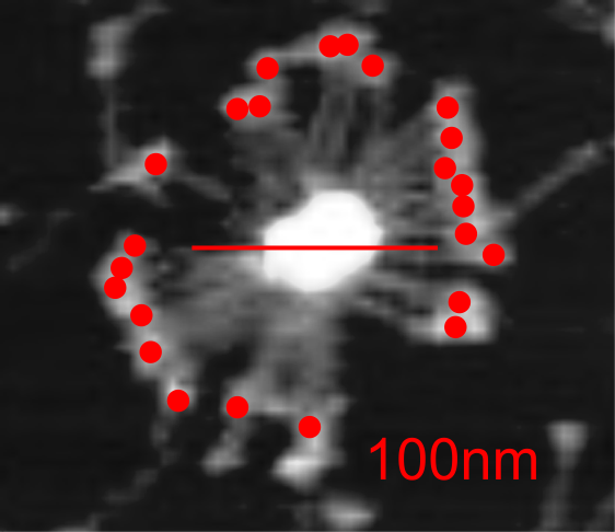
It is pretty obvious that the center of SP-D (at least as is represented in dozens and dozens of TEMs, and AFMs ) is longer in the direction of the 45 degree angle than the amino acid sequence would suggest and longer too than most diagrams present. There is a central dense region (which is found also on the tracing) but an extended region where two arms of the dodecamer are closely aligned with the combined length of something around 30nm, or a little less than a third of the entire length of the dodecamer.  The separation of the two arms on either end into the @45 degree angle  leaves the remainder of the arms as the collagen-like domain, and the neck and CRD on the order of 10-13nm the latter folded back upon the neck section to some degree. Tracing from Figure 2. Arroyo et al. JMB. 430, 1495-1509, 2018. Their measurement for the total width of a surfactant dodecamer seemed to be considerably larger than measured by others. So a compromise in estimated dimension has to be taken because of the differences in measurement accuracy and how much spreading of the molecules has occurred…  Relative measurements then, of 100 nm might be useful in determining the relative lengths of the segments of the dodecamer, but not the absolute lengths.

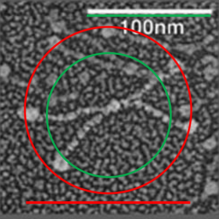
Normalizing the dodecamers to 100nm cutting centrally through at least two (probably best making it two polar) CRD is useful. A summary of the center (the highest, brightest (by TEM and AFM) areas of the SP-D dodecamer might be about


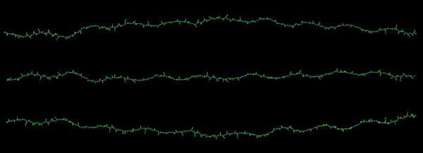
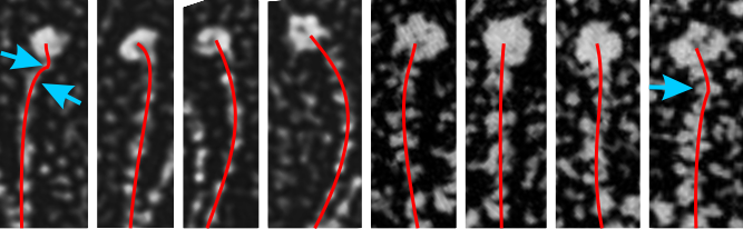
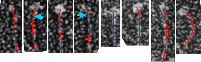
I would like to find a way to trace density of a curved line…. straight lines are no problem.  I think it is time to go back and measure these all one more time but setting density and width standards to get relative dimensions which at some point could be used for generalities describing proportions and regions involved in SP-D. s

Lost collagen domain length in mini-SP-D mutant

After figuring out that the images from one particular publication are between 15 and 20% larger than the micron marker accompanying the figure indicates, I went back to re-determine how much dodecamer arm length that the mini-SP-D lost when exons 3 and 4 were deleted from the collagen domain.  Again measuring the full SP-D in the micrograph and then using other images of SP-D and their respective bar markers I think that after several sets of measurements the miniSP-D has been reduced to about 35% of its original arm length. I am measuring the center portion (which surely contains more amino acids than just the N terminal) where it bifurcates.  Different sets of measurements sort of come in around 25 or 6 nm. The CRD are pretty close, and measurements indicate that the top left figure (which is the one which has the out of sorts micron marker (indicated by the different measurements for the diameter of the  underlying SP-D dodecamer… the red circle being what was given as 100nm (clearly too short) and the green circle indicating a diameter that would intersect the CRD in 3 different locations (typically I have used intersection of 2, but this one has three by chance) and is a much more accurate indicator of a 100nm.  Using the later, the center is N term plus whatever of the collagen portion is associatiated) is about 26 hm..  in the full length AND the mini-SP-D  so this suggests that the measurements are more in line, and also that the central X of the dodecamer hasn’t changed much with the deletion of the C3 and C4 regions.   Bars used to measure the arm length are the thin black lines in the lower right.


I am not putting these numbers out as anything but approximate, but they are similar to earlier posts, and a general indication of size.

One thing that is kind of interesting to me is that the collagen-like domain is often referred to as “disorganized” and often referred to as “braided” or like the helix of collagen.  There really should not be much discussion on whether it is organized or not… haha…  just looking at the pictures.


N terminal of mini SP-D dodecamer: is it different in shadowed images?

Just a thought here….which came to mind while i swas measuring the arm length and CRD dimensions and N term lenght of 11 mini-SP-D molecules that there was something odd in the center of about half of them. The N terminal of mini SP-D dodecamer (White et al, mentioned frequently) sometimes has an odd look bundled up central portion. I copied and pasted these 11 images to compare them, and 4 out of 11 really did have a greatly bundled (and seemingly) a reduced length. Typical full length SP-D makes a dodecamer with an N terminal (which might also include a portion of the collagen-like domain… as the proportions given in diagrams and in number of amino acids in the N term seem to disparate, and not aligned with the lengths seen in the shadowed electron micrographic images). In particular however, the mini-SP-D with deletions in the collagen like domain show N terminals (about 36% of the time) which are “balled up” into dense areas. Just looking at the layout of 11 molecules below it is also apparent that in those images where the N term areas is balled up, the collagen like sequence (arms) are separate earlier.

In two of the four dodecamers on the right, the CRD of an arm is not clearly defined, there is the possibility that in those cases, the CRD has fallen back on the N terminal. Rejecting that idea, there are also images in the left hand column which have poorly defined CRD and they do not show balled up N terminals.  The nicer explanation is that somehow removing the C 3 and C 4 alters the way the N terminals bind together in the center.

That the bar portion in the center of SP-D and mini-SP-D has some of the collagen-like sequence seems

i think had i been doing this microscopy i might well have been advised to be absolutely precise in my measures of nm length.  In retrospect in my own career…. i should also have been more careful.

What does the collagen like portion of SP-D look like?

Haha….  It is so clear from electron micrographs that the collagen like domain of SP-D is pretty much straight, not quite, arc of xx degrees but I am searching for anyone who has modeled that portion.  In Phyre2 (i do not know how long this link will be active). I found two linear models for structural protein which are pretty close and have a slight curvature (though these are single lengths not wound trimers so shape would change.  it seems to be a reasonable guess that the two are pretty much similar. Here three molecules with high similarity to the following sequence for SP-D as a “hint” of what it might look like.  The following is what I putinto Phyre2(>collagen_like_domain_SP-D
GLPGRDGRDGREGPRGEKGDPGLPGAAGQAGMPGQAGPVGPKGDNGSVGEPGPKGDTGPSGPPGPPGVPGPAGREGPLGKQGNIGPQGKPGPKGEAGPKGEVGAPGMQGSAGARGLAGPKGERGVPGERGVPGNTGAAGSAGAMGPQGSPGARGPPGLKGDKGIPGDKGAKGESGLP)

Three molecules rotated top to bottom, and i will try to weave them together in a helix.  Below that is the closest modeled sequence shown on Phyre2 from which the structures were saved in

Protein feature view i am wondering if this is more or less to scale.

the N terminal plus 8 repeats in the collagen-like domain were not well modeled. (>Nterm_SP-D_plus_8_gxy
MLLFLLSALVLLTQPLGYLE (signal peptide) AEMKTYSHRTMPSACTLVMCSSVES (N terminal) GLPGRDGRDGREGPRGEKGDPGLP……….. (first part of collagen like sequence)

Discrepancies in nm size of SP-D – more

Here is what i have figured out as a measure for the “real” magnification of panel A in the paper by White et al, 2008, on rat and mutant SP-D molecules.  My original observation was that the SP-D dodecamers were too large for their 100nm bar marker. That I attempted to confirm by creating two circles, one the size of their bar markers, the other a circle that intersected at least 2 of the CRD in the SP-D dodecamer.  The discrepancy was large, being about 132%.  I next could see that the background had a larger grain in A of their figure than B (and now i believe it is off even more in C) and measured that, and A was about 115% larger shadowing grain than B.  Thinking of different shadowing on different grids which must have some variation, I felt that could be improved upon, and surmised that the mutations in their mini SP-D was only in the collagen-like domain, not in the neck or CRD.  That said, I measured height and width of the CRD in panels A and B and compared the mean of those measures.  (there were 31 observations in panel A and 80 observations in panel B.  Some i didn’t measure as they were obscured by other details. The means and sd showed a significant increase in the size of the CRD in panel A over B.

Here is the micrograph, and the pink dotted lines show distances measured (a height and a width per CRD).  This too showed that A was about 115% enlarged over panel B. So now when one (as i will) measures the decrease in the collagen-like portion of the mutant (mini) SP-D, i will enlarge the images in mini SP-D in panel B before comparing them with the images from panel A.  That the neck and CRD in panel C are again smaller i wont work with this since there maybe some change (as observed in the “lack of separation” in some of the CRD in the images, so other things are changing the shapes there.  (please note that the background grain size is even smaller than in B. (so much hassle for such a simple error)
Thank you to two great websites which I totally love, as i can cut and paste from excel and get numbers i want.  SD calculator, and t-test calculator.

Discrepancies in nm size of SP-D

Here is where it become problematic when defining the size of anything as an absolute…. all these measures seen in publications need to be taken in a sense of wide variation in different reported sizes for the 100 nm micron marker.  Case in point, from White et al, their 100 nm marker (my green bar underneath their own white nm marker) and my green circle (the same length as the diameter as my green bar marker) are shown without changing anything on their image….. this molecule is at least a 132% over what most individuals report as the alleged size of the 100nm SP-D dodecamer molecule.  SO, if this rat SP-D dodecamer is that much bigger than the mini-SP-D which is claimed to be missing — “A rat mutant with deletion of two internal collagenous exons corresponding to exon 4 (C3) and exon 5 (C4) was generated by PCR overlap extension using full-length rat SP-D cDNA as primer”, how is one to know what the “real” size difference is.

Here is an image using their own bar marker (green) and the diameter bar marker (peach), showing the relative decrease in arm length…..  This is a 72% decrease from the rat SP-D in their own micrograph, and an additional decrease when using the “other researchers” estimates of 100 nm SP-D molecules.

in trying to figure out the discrepancy in size of the molecules of SP-D in this publication i decided to do just a simple test: to take background images from the full SP-D  and the SP-D dodecamers with the collagen portion deletions and see if they were anything similar in texture.  So this also begs the question of differences in shadowing techniques on different grids…  so that could be part of the problem but here clearly there is no similarity in the size of the shadow particles in the top (rat full length SP-D molecules) and the so called mini_SP-D dodecamers.  This makes me think the full length SP-D is not the magnification reported. From E. Crouch, et al 1994   “SP-D revealed a highly homogeneous population of molecules characterized by four or more relatively rigid-appearing subunits, non-randomly arrayed about a central core. The core was only poorly defined as the center of interaction for the N-termini of the subunits. The subunits seemed to emanate from opposite sides of the core in pairs closely apposed over a distance of 10 nm from the center of the core. The results for paired subunits presented here correspond very well to the structure of SP-D observed in electron micrographs. Site-directed mutagenesis of Cys-15 and Cys-20 has shown the necessity for N-terminal cysteine residues to stabilize oligomerization, but the cysteine residues are not necessary for trimer formation. However, other results provide strong evidence for the existence of at least one intra-subunit disulphide bond within the N-terminal region after limited peptic digestion.It is possible that apposing pairs of SP-D subunits are associated in an overlapping anti-parallel arrangement restricting the spatial distribution of the subunits around the central core. Another hypothesis is the association of paired subunits end to end. Two subunits are disulphide-bonded between and within subunits, whereas the two (or more) opposing pairs of subunits are associated through an undefined molecule. It has not been possible to exclude the possibility of the contribution of other molecules in the assembly of several SP-D subunits. It is also likely that cross-link formation between two or four subunits involves previously unpaired cysteine residue, or that a process of rearrangement converts a specific subset of intra-subunit disulfide bonds to inner subunit bonds.”  this is good in a sense that when the centers of the fuzzyballs are examined, there is not an N-end to N-end “look” about the assembly.

I am looking for the predicted length of the collagen-like domain with the C3 and C4 exons.

Total SP-D dodecamer measured length using arc angle dimensions

If i use the distance between two trimeric arms of SP-D seen in dodecamers as the off measured 100 nm width of the molecule, then measure the arc angle of each arm (arc angle length determined by [ 2pi R  (central angle/ 360) ]  this increases the total length (stretched as if a planimeter were used) of the separated collagen-like domain of the dodecamers at about 39nm for each half.  (n=14 individual dodecamers with n=4 arms each), or the mean of the lengths of the two sides as separate individual measures (n=58 *one dodecamer had what looked to be five arms and was included).

The mean dimensions in nm of the N terminal domain, taken as one measure probably includes the N terminal as well as some small portion of the collagen-like domain, still both tethered together tightly (that is, the N terminal and a portion of collagen-like domain) and is about 19nm. The measurement spanned the area between bifurcation of the collagen-like segments on either end of the “joined” N terminal domains. (purple line)

The CRD measures 10.8nm whether determined by the mean of the length of the two sides, sorted by dodecamer (n=14 dodecamers, each with 4 CRD), or the mean of the lengths of the two sides all as individual measures (n=58 *one dodecamer had what looked to be five arms and CRD and was included).

A total dimension of about 119nm (the individual image shows a total dimension of 112.7nm – below). The very same diagram is shown above and below, one made before i decided to check how to measure the curvature as part of the length of the trimeric arms, the other after.

Here is the image posted earlier, where the arc length is NOT calculated, but just the straight line distance between where the trimeric arm diverges visibly from the center (Nterm plus whatever collagen-like sequences are there).  When measured in parts, the total length of the molecul below measured at 107nm slightly above the 100nm which I constructed as the diameter between two CRD lying on a the circumference of a 100nm circle.

What is pretty clear is that there are not that many, if any, accurate diagrams out there that have the proportions of CRD to thickness of the collagen like portion, or the curvature, or the length of the four segments of SP-D right.

One thing for certain, is that shadowed and AFM images of SP-D show that the collagen portion is certainly NOT straight, as referred to in the chapter Alveolar Epithelium and Plumonary Surfactant, by Robert J. Mason MD and Leland G. Dobbs MD in the textbook Murray and Nadel’s Textbook of Respiratory Medicine, 8, 134-149.e5.

I could continue measuring all the dodecamer images i have collected using the arc length, central arc angle, N terminal (and whatever else) bound central segment, and size of the CRD but i don’t think adding those new data is going to change the outcome.

Here is a mutant surfactant protein D fuzzyball (from Kevan L Hartshorn, et al – with the mutation in the CRD so not likely affecting the overall dimensions much) which is greater than their own micron marker would designate as 100nm. This image was cut and pasted into photoshop from their figure, a dupllicate made, top layer increased in contrast, decreased in brightness, and layered at 50% transparency over their original in order to show the arms of the dodecamer and the CRD better. This is a mutant SP-D, according to them, with two substitutions in the CRD. (Recombinant human SP-D having a Met at amino acid 11 (and Ala at amino acid 160).

Counted the CRD just for the fun of it.

Arm kinks in surfactant protein D dodecamers

Surfactant protein D is a terrific molecule, and the assorted arrangements of multimers has been described somewhat but I have only found one publication which actually tries to figure out details about the unique shape of the collagen-like domain. The carbohydrate recognition domain and the coiled-coil neck section has been studied – ad nauseum and the RCSB has countless molecular models. The N-terminal has been a little less studied, even less the collagen-like domain.  I am not really sure why that is since there are dozens of images of SP-D trimers, dodecamers and multimers (fuzzyballs, fuzzy balls) from which to determine length and curvature of that domain.  At anyrate, there are some images which show kinks at the neck (a comment typically included in descriptions of SP-D, so that is not new) and at least one paper suggests there might be something going on at the glycosylation site (closer to the N terminal).  Here is a group of just for trimeric arms (cut and rearranged from dodecamers (images from manuscripts that I did NOT write – thank you for those) and it becomes clear that an assessment of angles in this kind of order might be useful. One kink, maybe two kinks (see blue arrows on left).

It is pretty clear that the length of the collagen-like segment that show up right at the crotch of the bifurcation of the arms of the dodecamer is not going to end up the exactly length of the whole collagen-like portion. It seems at this point that the there is N terminal association of the four trimers and then more close binding for a greater length up the collagen-like sequence.  The proportions of the micrographs just don’t add up (especially visually) to the dimensions given in the literature for the collagen-like sequence. In particular too, the diagrams are way off. This begs the question, where does the central ly bound portion end? and does this make a difference in the shape of the center of the fuzzyball made up of 6 or 8 dodecamers? as i mentioned weeks ago on this blog.