It is worth reading this nice review article which deals with adherens junctions and also some other cell adhesion molecules.
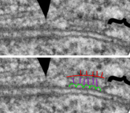
Oda and Takeichi 2011 have this nice electron micrograph or cell-cell adhesion, but it is even more interesting to me that the area described with the confines of their arrows, and the area within their bracket, does not show as distinct an organization as the area inbetween their designated zones… Haha. see bottom two micrographs, their photo unretouched, and the area where I see amazing order, an area which is very organized.
semicircular arrangement (black lines), likely proteins which attach to whatever membrane proteins the circles are, and then the red lines as some molecule which links the former with some cytoskeletal element. There is a great microtubule over by their arrows, i don’t know what is under their bracket.

 Just a note: it looks like the primitive junctional complexes in drosophila are the result of one-size-fits-all type molecule with little segments snipped out as DE-cadherin has every thing but the kitchen sink there….including laminin, proteolytic, and EGF-like domains.
