Category Archives: Lichen planus

Pathology report: lichen planus

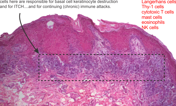
Well, that was slightly disappointing. Looking for, at most 6 minutes, through three slides was less than uplifting. The classic look: mild hyperkeratosis (thickening of the stratum corneum, or that uppermost layer of skin cells (which are without nuclei) and usually includes some additional keratin; infiltrate and apoptotic basal keratincytes; and acantholysis (the loss of intercellular connections, such as desmosomes and adhesion molecules (that can cause separation of keratinocytes in the middle and basal layers, and also signal cells to undergo apoptosis) and lots of cell debris and apoptotic bodies in the basal layer of the skin. With this there are numerous infiltrating lymphocytes (which the literature calls Th-1 cells) that apparently are able to send self destruct messages to the basal keratincytes. All this was surely evident in the slides (3 separate biopsies) from my own case of lichen planus. That said…. you ask, why are you disappointed?

Disappointed in the lack of “interest” in the real histology..like actually viewing at a magnification worth looking at… ha ha…. for an electron microscopist to be satisfied looking at 10, 40 or 60X H&E is like asking the driver of a Koenigsegg Agera RS to be happy on a scooter. While I recognize acutely (bad pun for a chronic dermatosis) how difficult it is to get through a day of reading slides I just didn’t get the feeling of thoroughness from this man, and also got a definite thumbs down when I suggested that there might be an environmental component. What gives? do the old MDs think that all of life grew up in a space-vacuum… the environment is critical.

Another question that I asked and was surprised at the answer was whether there was a good database for lichen planus cases…. he asked “database” So i asked again, to see if there were a lot of cases….so apparently there are not.

Online publications seem to suggest that lichen planus (i assume they include all variations) is 1-5% of the population? To me… that would be a lot.
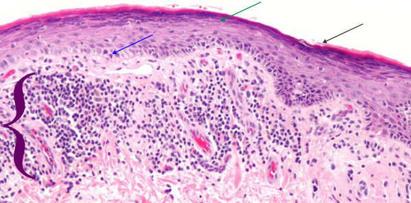
Here is a picture (wikimedia commons) which is a lot like (not exactly) what my slides showed.  Blue arrow points to keratinocytes which are undergoing apoptosis (sometimes by the old guys called Civatte bodies), green-blue arrow points to area of stratum corneum, and black arrow to the keratin layer.  White space is separation of intercellular junctions and are part of the disease process. Purple bracket = the band of infiltrating cells (for all practical purposes at this low magnification… one has to just call them mononuclear cells (which would include most cell types… ha ha). Bright pink rounded areas in dermis are blood vessels.

When i asked about langerhans cells, Thy-1 cells, melanocytes, that just drew a “no comment look” for obvious reasons, the magnification was too low, as was the interest in finding something out about these participating cells and the extent to which they might be found in different numbers in cases with different origins.  While I dont discredit the authenticity of the diagnosis from the pathologist i met with today, I do feel that the barest minimum of reporting was made, with great emphasis on “i need to move on to read other slides” and “dont bother me lady”.   This is part and parcel for being an old lady in science. It is ok to be an old male in science…. but the gender bias is alive and well.

THere was also no interest expressed in doing anything in the way of a study about the variety of immune cells, their possible connection with cumulative, and concurrent environmental triggers.  Ok, that makes me pretty energized to figure this out some other way…the pathologists here are not going to be of much help.

I have three new ideas:

I) the disruption of adhesion molecules (that would be dozens of molecules which comprise portions of the desmosomal, adherens and tight junctions, of which many participate in cell signaling i.e. apoptosis.  The adjesion molecules may be a direct or indirect response so that I will have to google.

2) The apoptotic cells: are these self induced apoptoses due to loss of adhesion molecules, or are these apoptoses  achieved through the infiltration of Th-1 cells (cytotoxic T-lymphocytes (CD8+) and NK (“educated”by langerhans cells)  which have as their goal to annihilate basal keratinocytes. A few ways to kill basal keratinocytes –by the release of perforin and granzyme or by the Fas/FasL system.

3) Are mast cells and eosinophils involved, particularly in the release of vasoactive substances, upon touching, sheering or stretching the lesions, which then triggers enormous itching.

4) another question would be, if this is indeed a “educated” set of Th-1 and NK cells, how long would one suppose that the “commitment” of those cells last.  Would it be a life long immunity gained, or something that passes quickly.
Eosinophils in lichenoid drug-induced eruption vs lichen planus

Good information to know in the differential diagnosis between lichen planus and lichenoid drug-induced eruption is the % of eosinophils.  That would be interesting. I guess I have to assume that no one in the derm path here at UC is going to make any eosinophil counts on my three slides.. ha ha. I would have to say, with H&E sections, eosinophils can stand out as cells with bright granules. I think mr. derm path that i looked at slides with yesterday would have had to up the magnification to see if this were so.  I am interested because it would increase the likelihood that my diagnosis was cement, concrete, mortar, grout related.  Publication with that data linked to yesterdays post and to this article here.
For me this is exciting information since differential counts of my peripheral blood smears has indicated an elevated eosinophil count (meaning various things, including a susceptibility to allergies) since I can remember… (so a long long time ago). This would make the diagnosis of my lichen planus more like lichenoid drug-induced (also called lichenoid interface dermatitis) for several reasons:
1) most serious lesions on shins – lower extremities, and now coming on the backs of my hands (abrasion from the leashes of two dogs when we walk, ha ha)
2) WHAT NEEDS TO BE EXAMINED in my slides…. the presence of 8-10 eosinophils per 20x field in the regions where lymphocytes are most dense. Gee… you would think that derm path could do that in about 10 minutes…. should i ask, or should i wilt back into oblivion because there is gender bias among scientists and mr derm path examiner himself would not be able to accept the challenge.

Anyway…. the point here is to make some connection between the lichen planus drug-induced (which in my case could be cement, concrete, thinset, grout-induced) dermatitis. Just thinking outloud here.

Review article: lichen planus

This article looks to be a reasonably comprehensive assessment of a disease that is varied and under-reported and annoying (i know from experience). Establishing facts and organizing data is about all we can do.
My goal is to try to figure out what the mechanism is so that I can contribute to an appropriate treatment option (selfishly for myself but hopefully benefiting others). An update on the food sorting and avoidance, I figured that after three or four weeks of not eating some foods, that if i did not see an improvement, yet even saw increase numbers of lesions that “food” was the least of my worries. But, that said, I am certainly increasing by volumes the amount of vitamin A that i am ingesting through natural sources…which leads me to recommend sweet potatoes, and red bell peppers, some dairy, since these foods have reasonable amounts of vitamin A. I am not inclined to eat organ meat, i could try fish oil.

Here is a tiny tidbit of data: When i had three biopsies (shoulder, leg, and dorsal forearm, a bandaid was put on each, and while NO lichen planus lesions grew in the biopsies…haha… they did appear under the tape marks of the bandaid. Now when Dr. Blabla says there is NO ENVIRONMENTAL component… they are just wrong.

That lesions are bilateral, mmm possibly, in my case it seems right side shins are worse than left… but so were the initial lesions created by my lawnmower (those pesky rocks, sticks, and shards of acorn) thrown back into my shins — the origin of the large lesions, and the only place that I have lesions larger than about 7-8mm diameter. Half a dozen of those on my shins are 20-30mm in diameter and 5-7mm thick. I think it is hypertrophic vesiculobullous lichen planus… two in one — LOL. I can confirm that the worst lesions are on “sun-exposed” areas of legs and arms.

This review (linked) above unfortunately does not do a good job of explaining all the nuances of the pathological architecture, structure, or ultrastructure and parrots what has been published in countless other places… though i suppose this is due in part not having a truly committed pathologist. I have tried but have not been successful (even though promised by the MD) to have access to the slides to read them myself.
When i read the slides i will take careful count of the mast cells, eosinophils and basal keratinocyte destruction to figure out if this (my case) is a lichenoid drug/chemical (in my case would be chemical) reaction rather than classical cutaneous lichen planus.
In retrospect, those who name the disease for the look of “lilchen” on the barks of trees sort of did the whole deal a disservice. And while they did not know the cause, and the cause is not known today, might have been smart not to name the disease after something that grows on tree bark. ha ha

The list of drugs which cause lichen planus like lesions is long and the number of drugs on the list given in this report would include something taken by almost everyone, and includes simple things like ibuprophen. good grief.
One thing that keeps coming to mind is perhaps a mixed-cause… that underlying condition possibly left over from a shingles outbreak, or a viral infection, or maybe even a series of bug bites that stimulate some immune response that is triggered to go into overdrive by an environmental exposure (as in my case, perhaps related to mortar, cement and ground exposure over a 10 year period). Would that someone in this insitution (there are those, like the Berensteins here in the department that are in tune to environmentally and occupationally triggered diseases, but they were too busy to take note. Just to belabor the point, the university of cincinnati does NOT take care of its own. One would think that being 50 years at this institution that I could call upon one of the MDs, (someone in the same department of environmental health, as i am, like the Bernsteins) and expect some kind of compassionate treatment…or to be able to email the pathologist who was supposed to have looked at the histological preps, Dr. Mutasim) but no such luck, in fact quite the opposite — how sad). NO perks for having spent 50 years doing research here.
Paper listed above does regard many metals as possible factors in LP reaction…so if the metals can trigger this in crowns and fillings, why not also metals found in other occupations, e.g. cement, mortar, grout, tile and similar applications. Still i could not get the attending MD in my case to do anything but scoff at the idea.

AND HERE — my fav line in this paper: LP is a complex disease and thus can be caused or triggered by genetic malfunction and/or environmental factors.

For what it is worth (for me it was worth much) here is a pdf file that some of you may be able to read and find helpful. For me the help came in this paragraph ”
The high proportion of cases reported among these populations suggests a genetic susceptibility for LP. This idea is further supported by cases of familial LP, reported in 1 to 4.3% of childhood LP series (Kanwar and De, 2010; Nanda et al., 2001). However, familial cases have not been reported in all series, and specific antigens have yet to be determined. Pathogenicity of LP lesions involves the autoimmune-mediated lysis of basal keratinocytes by CD8+ lymphocytes, though definitive etiological triggers are still unknown. Case reports in both adults and children have shown an association between LP and the following: chronic liver diseases such as chronic active hepatitis (particularly hepatitis C) and primary biliary cirrhosis; complication of hepatitis B vaccination; viral and bacterial antigens; trauma (via the Koebner phenomenon); metal ions; medications; and a variety of autoimmune diseases such as autoimmune thyroiditis, myasthenia gravis, alopecia areata, vitiligo, thymoma, and autoimmune polyendocrinopathy (Luis-Montoya et al., 2005; Pandhi et al., 2014).  IN PARTICULAR  linking METAL ions to this disease is what I am thinking happened in my case, the metal ions as chromatic agenets in the thinset mortar and the grout that I used on my basement floor. Chromium and cobalt, and hopefully no cadmium, but plenty of other metal elements, and also including the silica, known to cause respiratory disease.

This particular paper suggests that corticosteroids are beneficial, but many sites and publications do not give steroids much credit for shortening the duration of the disease, which can be years. I for one wasnt helped by topical steroid cream (betamethasone was the only one prescribed for me, outside of a very outdated half used tiny sample tube of topicort none others were used).

Next chapter begins a fun 11 or 12 years making ceramic tile collages, montages, outdoor pots (big big ones) and laying 35 thousand tesserae on my basement floor.  Little by little over that span i began to notice a dry cough.  Not that bad, but bothersome.

Case Report: lichen planus – which hypersensitivity? Chapter 2.

[aside]I dont mind posting the names of 5 MDs, none of whom were helpful. Dr Wones, Dr. Zubritsky, Dr. Carter, and Dr. S…(from the middle east) cant remember his name, and the pathologist Dr. Mutasim.  None knew anything about this disease, nor were helpful in any way, in fact Dr. S was downright derogatory, showing complete ignorance about how great the impact of environmental stimuli can be on diseases in general…that is pretty much an old-school mentality, maybe some “updated” information would benefit his overall knowledge of diseases. One sent me a link to a website about lichen planus which only had the most superficial information, totally useless. I guess he thought he was being helpful.

BTW i think that i have had the tendency for lichen planus for many years so what caused the explosion of lesions is really what I want to know.

Several kinds of hypersensitivity have been described (more will likely be found). TYPES I to IV.

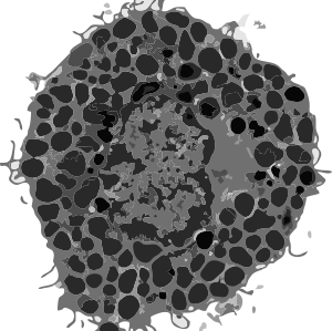
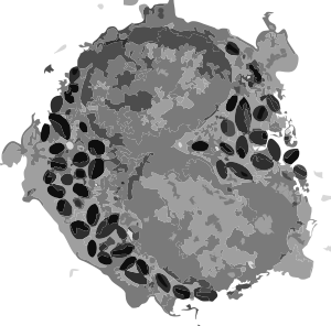
TYPE I -Immediate hypersensitivity happens fast. CD4 T (thymic lymphocyt) cells are involved in adaptive immunity (to pathogens), autoimmunity, asthma, allergic and tumor immunity. Naive CD4 T cells may differentiate into one of several lineages of thymocyte helper cells, (Th1, Th2, Th17, iTreg – about which I know nothing). After the first exposure to the allergen, the CD4 T cells are “sensitized” and immediately effect a response by releasing antibodies (immunoglobulin E (IgE))  which signal mast cells and basophils (with high affinity IgE receptors on their cell membranes and loaded to the hilt with storage granules) to dump out vasoactive amines, e.g. histamine. These chemicals then affect surrounding cells, mainly causing blood vessel dilation and muscle contraction, and cause the inflammation by increasing the permeability of the capillaries to white blood cells and some proteins (thus redness swelling and itching).  Histamine (one of the vasoactive amines) is a cute little molecule (right)(thankUwikipedia). Basophils (left) and eosinophils (center) are cute too (I vectorized two transmission electron micrographs here).

Delayed hypersensitivity is just that, delayed onset. Appearing several hours, days or continuing to appear a week or so after contact with the antigen and depending upon the dose of the antigen. For example poison ivy appearance (at least in my case) comes up between 2 days and a week after exposure, sometimes showing the secondary contact point of the original molecules of urushiol being where two skin surfaces meet..the original spot depositing a lesser amount of urushiol at the point of contact.  While i have never read about it, I do believe there can be systemic poison ivy, for instance when the neighbor weed-wacks their yard full of poison ivy and the droplets of urushiol are aerosolized and breathed.  Would be fun to search that route of exposure (in the literature, not as a test subject).

Delayed hypersensitivity is not antibody dependent, but is a cell-mediated response.


Case Report: lichen planus – is chromium an issue? Chapter 1.

I am going to document what I am thinking about while keeping my hands off the itchy places (not so LOL) to try to figure out what environmental+immune conjunction caused this to surface in my otherwise really healthy 74 year old body.

I don’t know if it will help anyone else in the search for cause, but hopefully it will, and it will allow me do write a history of my own experience (noting everthing I can remember and blaming no one and no thing or even being disparaging..well except maybe a little to the MDs that we depend upon to help us through diseases..and who (in my case) did “not much”. Ha Ha.

I am also going to encourage anyone out there to write a similar history, and though I don’t take comments on this website because so many unscrupulous individuals want to spam and skim and link for their own advantage instead of participating in meaningful discussion. My email can be discovered from the very first entry on this blog, way back when!!, and from one of those websites you can contact me if you want to contribute a long history. My goal is to treat this exactly like a manuscript, and submit it at some point to a scientific journal.

Starting at the beginning (not the beginning of my life obviously, that was 1943) but of contact dermatitis which happened to the best of my recollection in 1968 or 1969, a new time resident of cincinnati, and during a frisby throwing game in burnet woods.  If you are from cincinnati you know burnet woods is a primitive wooded area, small, more a park with a small lake and lots and lots of poison ivy.  I didn’t really know what poison ivy looked like, but in my leather sandals (imported) apparently I ground some poison ivy oil and got a really bad case of it from that afternoon.  I mention it because of the inflammatory process of the poison ivy itself, but back then, leather, and imported leather was tanned with some pretty harsh chemicals, including chromium.  Every time (which was probably one or two more times) I wore the sandals i re-got poison ivy.  Chromium is the key word. Urushiol is the next key word as it is the oil in poison ivy that causes the allergic dermatitis. HERE is a lay website for some important information. A host of things can cause allergic dermatitis; food, cosmetics, metals (i could never wear stainless, nickel plated or silver earrings) and it also can be caused by paints and mortars and cement and grout.

That the exposure on my feet was to 1) several unknown tanning compounds, 2)as well as the urushiol (which causes a T-lymphocyte response), what appears to me to be a really good combination for an adjuvant effect, causing an senhanced response.

I did get poison ivy pretty seriously in 1975, but to my knowledge there was no other environmental exposure at that time. (except possibly, chromium in silk screen paint which i used a lot of (and probably still have some in my basement… SKRINK brand)..greens and blues.  Direct contact of Cr(VI) compounds with intact skin can induce chromium dermatitis or sensitization. and a government document on chromium toxicity.
So chromium has been a part of my life for a long time, though maybe not in a good way.

Fast forward many years, to “fun with polymer clay” when in the 1980s i used lots of FIMO, and likely all colors, though I cannot distinctly remember which colors i used most, but very definitely there was blue, and likely green.  I used enough of that early FIMO to produce a contact dermatitis in the palms of my hands, little bumps, not terribly itchy that i can remember, but enough to make me “wake up” and know that polymer clay in that formulation was a contact allergen for me.  And fast forward another 30 years and it is back to polymer clay again, not too careful this trip around, but no contact dermatitis on my hands (the clay does stick to hands…. especially with some pigments,  black clay and red polymer clay are particularly noticeable, but I don’t doubt for a second that they all stick.

The take home message is that we do get exposures…to many things…. not all of which our bodies treat as benign.


Lichens planus: my thoughts!

BEST post i have seen by far comes from osteopaths…. that says something about this group.
and then i read the American Skin Association’s site and it looks pretty much the same… go figure.  Lichen planus is a relatively common skin disease that comes in episodes lasting months to years. The onset may be gradual or quick, but its cause, like many skin diseases, is unknown. It appears to be a reaction in response to more than one provoking factor. Theories include stress, genetics, infective (viral hepatitis C) and immunologic (autoimmune). There are also drugs that produce lichen planus-like allergic reactions to high blood pressure, heart disease, and arthritis medications. There is an inherited form also which is often more severe and can have a protracted course.

Lichen planus appears as shiny, flat-topped bumps that often have an angular shape. These bumps have a reddish-purplish color with a shiny cast due to a very fine scale. The disease can occur anywhere on the skin, but often favors the inside of the wrists and ankles, the lower legs, back, and neck. The mouth, genital region, hair and nails are affected in some individuals. Thick patches may occur, especially on the shins. Blisters may rarely occur. Bumps may appear in areas of trauma on some individuals. About 20 percent of the time lichen planus of the skin causes minimal symptoms and needs no treatment. However, in many cases the itching can be constant and intense.

This disease occurs most often in men and women between the ages of 30 and 70 years. It is uncommon in the very young and elderly. All racial groups seem susceptible to lichen planus.

There is no known cure for skin lichen planus, but treatment is often effective in relieving itching and improving the appearance of the rash until it goes away. Since every case of lichen planus is different, no one treatment does the job. Topical corticosteroids are very useful. Antihistamines may be prescribed to relieve itching. Extensive cases may require the use of oral corticosteroid (cortisone, prednisone) for a number of weeks. This usually shortens the duration of the outbreak. For severe cases powerful treatments include photo chemotherapy light treatment (PUVA), the retinoids drugs (Soriatane and Accutane), cyclosporine and hydroxychloroquine.

Other helpful measures include soothing baths (Aveeno Colloidal Oatmeal, Nutrasoothe) and the application of wet dressings (tap water, Burows solution 1:40) to the affected areas to help reduce itching. Also, the use of lotions containing anti-itch ingredients such as menthol, pramoxine and phenol (Sarna, Aveeno cream, Prax, Itch-X) may be helpful.

As it heals, lichen planus often leaves a dark brown discoloration of the skin. Like the bumps themselves, these stains may eventually fade with time without treatment. About one out of five people will have a second attack of lichen planus.

for those of you out there (like me) that have the misfortune to experience this….. blessings to you.
I was not very impressed with the general practitioner, the first resident dermatologist, second resident dermatologist, or the attending staff that I have seen about this, NOW DIAGNOSED BY MICROSCOPY, disorder. I am calling it a disorder because to me a disease is caused by an infectious agent. Diseases and disorders both may have known and unknown causes. A disorder in my mind is more a “out of order function” which may be caused by an agent (any environmental influence) but the basic processes involved in the reaction are normal processes gone ary. Compare, bacterial endotoxins, or agents that cause infections like viruses and bacteria,
From what I have read lichens planus resembles some excellerated immune response. Histologically the presence of Langerhans cells, lymphocytes, changes in the mitotic rates of the basal and lower levels of the epidermis. I even think that what I have been reading about lately (cell adhesion molecules) might be involved since not only does the epidermal layer of the skin (in lichen planus) look different, but several mentions of decreased desmosomes between/among keratinocytes occurred. This could be related directly or indirectly to the disorderly processes of this condition.

Dont bother googling the pictures for this disorder… ha ha…. too scary.