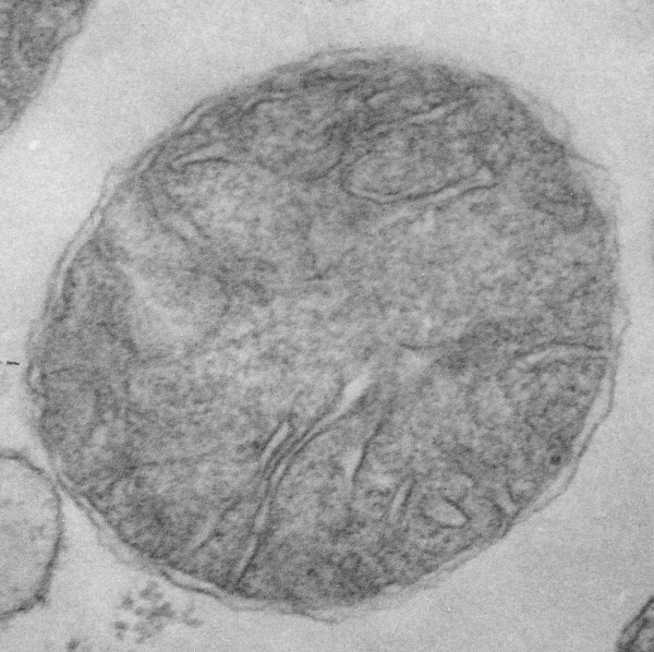
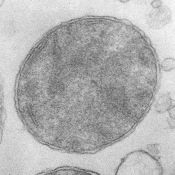
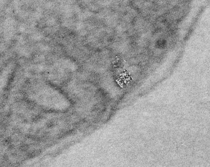
Such a huge number of mitochondrial features remain to be analyzed in each of the studies done in the past. Micrographs are available, but few choose to revisit those experiments that were done with care and integrity…for clues that verify or refute the claims made by others about such anatomical structures as mitochondrial pores, the position of mitochondrial ribosomes, the circular mtDNA and oligomerization of proteins from the respiratory chain and or oligomerizations of Bac and Bak apoptotic proteins on the outer membrane. So beginning with wild type (controls for a GCLC ko mouse study (which has been published – so experimental methods are available online) I will just look at the morphology of mitochondria in images already collected. 20,000x pole piece V, enlarged 4 x, neg 18322, block 78149, postnatal day 14 mouse liver, +/+ single isolated mitochondrion taken from the lowest section of a pellet of isolated mitochondria 6 21 04. (two mitochondria from the same micrograph, not identical enlargements)
 here is a snapshot of the lower right hand corner of the above picture with two mitochondrial RNA models superimposed at approximate scale, the one on the left, yeast, the other ? maybe mammalian.

