Monthly Archives: April 2018

Mitochondrial inclusion: matrix proteins?

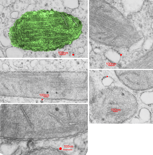
Hunting for an answer to what might be the inclusions seen in mitochondria in my micrographs from Rhesus monkey liver I ran onto this awesome paracrystaline inclusion found in mitochondria in some myopathies. I have searched and really have not found a great micrograph, but several pretty nice ones, and am reproducing what looks like an oligomerization pattern to these inclusions.

1) they don’t appear to be formed in cristi but more in the matrix.
2) it seems like there is a limit to the pattern of oligomers – and a border
3) different micrographs show different orientations of the patterning

Cut and paste portions with reasonable resolution and partial diagram of patterns. It is not really clear from micrographs available that these are matrix or intracristi..but they do seem to have a bounding membrane. Top row of segments look like three layers: row 1 flat; row 2 staggered 45 degree end on, row 3 staggered flat.  Middle row looks much different with less than a 45 degree angle to the middle row, and a longer and shorter component, and for rows 1 and 3, a taper to the “bricks”.  Last picture on the bottom right… different again.
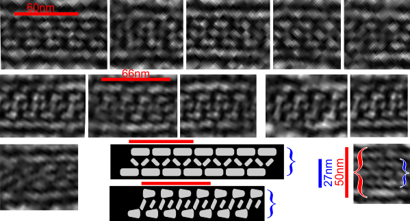
Mitochondrial inclusions

Liver from a rhesus monkey shows some mitochondrial inclusions.I found creatine kinase inclusions – paracrystaline but they appeared to be continuous with cristi membranes, SO MAYBE this (as found by someone else below) is not a perfect match but certainly a better match than a summary of the mtDNA mutations which are even more awesome than the paracrystaline inclusions in the monkey liver here. (anm# 97A mid lob, liver, control biopsy december 1974, negative 5743, block 4360, modified karnovsky’s, Epon 812 – to the best of my knowledge).

I havn’t included all the links, but ONE link i am adding which was an awesome summary of different paracrystaline and concentric cristi arrangements. FIND THAT ARTICLE LINK HERE. Mitochondrial inclusions, in general, would be a great topic for study, relating them to various over production of proteins, oligomerization, and loss of function.

YES: GRAVITY is part of the environment

How awesome is this study?  Ha ha…. having been in environmental research for 4 decades, never once did i consider looking at cells without the affect of gravity, and not to make a really bad pun, the gravity of the loss of gravity. FIND THE ARTICLE HERE. This group presents some great micrographs of the nucleolus….  I cant wait to see if the non-bilateral division of the nucleolus in one of their figures can be expanded to the very definite bilateral organization of the nucleus in general which is seen upon preparation for mitosis.

Perfluorooctylbromide: in rabbit hepatocyte

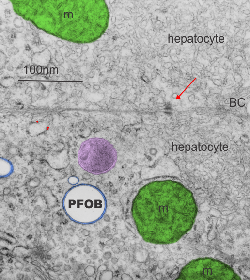
Rabbit liver, two hepatocytes revealed in this electron micrograph. Three possible, one definite, inclusion left over from the infusion of perfluorocarbon, from my notes it looks like perfluorooctylbromide (19.4%PFOB 5%F68), sacrifice dates show that it was more than one year affter infusion.  The mitochondria are pseudocolored green, the microbody purplish, the PFOB inclusion complete round (the density of PFOB is greater than water, which I surmise is the reason these structural footprints are completely round and empty) and the  very tidy layer of proteins around the perfluorocarbon droplet and bounding membrane are blue.  A small (less than 100nm) droplet is typical for the remnants of infusion of the more desirable perfluoochemical blood substitutes. This particular droplet does not show a “black cap” of enzymes which often accompany the droplets. Nice little desmosome between these cells and a bile canaliculus barely seen on the right hand middle.  Golgi in the lower cell at bottom with another microbody (peroxisome – which i should have colored as well.  My recollection is that infusion of some perfluorocarbons was accompanited by proliferation of peroxisosomess (Neg 5890_block_4783_28000x.

Mitochondria: parietal cells are mitochondria – rich

Mitochondria: parietal cells are mitochondria – rich as I see this counting up numbers of these organelles in cells while also tabulating the number of mitochondria which are tethered to desmosomes.  They are large, fat and everywhere, occupying much of the cytoplasm (as opposed to other cells where there are secretory granules or RER and ER machinery. They are quite close to all plasmalemma designations,  e.g. apical, lateral and basal, and even the canalular membranes BUT leaving a consistent small critical space between them.

Just adding a note, they really are sparsely populated with desmosomes and also with desmosomal-mitochondrial tethers — which is surprising i think and out of thosands of mitochondria counted in parietal cells only a few are tethered to desmosomes on the lateral membranes of parietal cells.

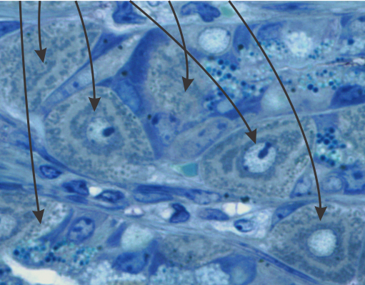
In this toluidine blue stained plastic 1 micron thick section you can see the darkish greenish dots around the pale looking canaliculi of parietal cells (arrows) nested closely and packed between the membrane structures.


Young Sheldon: april 19 2018 episode

If i had a rating scale for TV serials  i would rate Young Sheldon way up there at the top with a 10 if the scale is 1-5 (haha). It is so full of great language, great puns, great human assessment, and dilemmas that it tickles the soul and lifts the spirit.  Last nights episode with guest star Wallace Shawn as an eccentric old physicist, but not to old to find Mema disarming was a treasure that made me smile, and laugh, and re-think it again and again. Iain Armitage is a terrific little actor.

It would be wonderful if all life’s problems could be this amusing.

Apoptotic nucleus

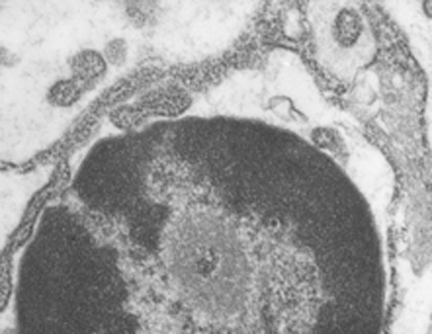
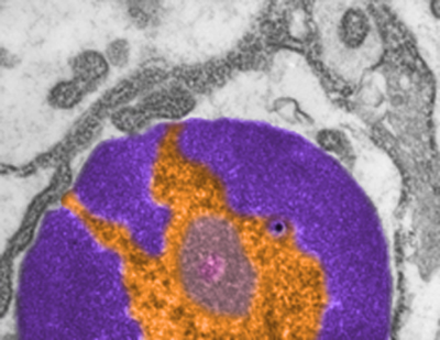
I wish i had taken a higher mag picture of this apoptotic nucleus as well as identified the cell type. I do know it is from animal #500 in a study of aged ghkaα-/- mice, this particular KO was 12 months old. It could be a parietal cell since in other image parietal cells are shown to have a “glassy protein” (stains without much texture and eosinophilic with H&E) (just recollection here). The RER is greatly, ribosomes are very closely packed (indicating the production of large amounts of possibly large protein products. Checking the fairly electron lucent contents of the RER there doesn’t seem to be any obvious oligomerization, polymerization or layering of the contents.  The light areas in the perinuclear space and around is dilated RER. It could also be a zymogen secreting cell.
One nuclear pore on the middle left nuclear membrane, and a part of another to the right and above that one. Packed ribosomes are in array on the RER membranes (the black and white cytoplasm pictured around the nucleus),  lots of condensed chromatin (purple) which may be exaggerated by the section being somewhat tangential to the widest diameter of the nucleus, and adjacent euchromatin (gold) which in this case looks pretty dark (not just because i pseudocolored it).  There is a prominent perichromatin granule just to the lower right of center (black) and interchromatin group (sometimes called nuclear speckles which to me makes no sense) is light violet.  Pinkish spot in the interchromatin granule  is probably just euchromatin?

Annulus around desmosome – freeze fracture

Here is an image from a letter to the editor (which i didn’t read thoroughly but was just looking closely at the freeze fracture of tight junction encircling a desmosome) and saw what I had seen in the transmission micrographs of thin sections: that is…. a small ring around the desmosome which i called the annulus which is restrictive and different than the intercellular space nearby.   This particular freeze fracture micrograph (original found here)  by Yasuo Kitajima shows it clearly, yet is it not marked. I don’t know what the function is but clearly a “no fly zone” surrounds the desmosome. Top micrograph untouched, bottom micrograph with bar marker and blue dotted lines showing part of the annulus. It is kind of nice that this particular desmosome is approximately what the literature suggests in terms of number of nm for the diameter.


so, next question is what are the semiregularly raised areas…?


Just because it says gluten free – organic doesn’t make it food

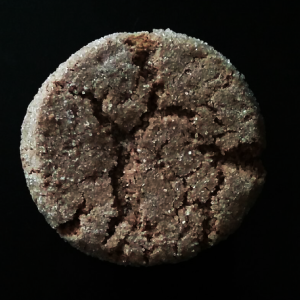
Just because it says gluten free – organic doesn’t make it food!  This nice looking ginger cookie, but a rather expensive cookie at that, because at regular price it is about 1.25  a hefty price for a single cookie.  I bought it when it was down to a quarter, in the WHOOPS we backed too much bin… even then… I could have bought a lot of white sugar for that sale 24c and that is about all that was in this cookie. I could not believe how much it tasted like eating a teaspoon of white granulated sugar. The texture was totally like granulated sugar.

This is not food people…. this is marketing and hype.

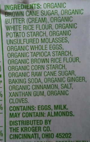
1 cookie, half fat, and at that a whopping 14% of my total daily saturated fat. list of ingretients…. first organic brown cane sugar….  ha ha…  no surprise there, and organic sugar will give you type II diabetes just as fast as non-organic sugar. 17 (give or take one) ingredients, and 7 are sugar salt and butter.  I might be mistaken (not) but putting the label “simple truth” on a package, and adding gluten-free, and non GMO and organic doesn’t mean anyone is selling you a good thing to eat.  I just means they don’t mind using buzz words to get you to purchase stuff and confuse your conscience.