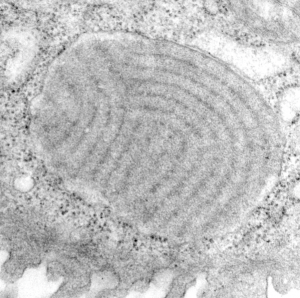
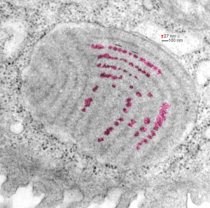
Alveolar type II cell layered granule in ferret: even the dense bands have periodicity. I had generally thought that the outer dense bands of the layering in alveolar type II cell granules of the ferret were pretty continuous. It was easily evident that the middle band in each period of the multilayered granules displayed a periodicity (5-7 per 100 nm), but I had not seen much periodicity in the more dense outer layers of each period. On several tangential sections of these granules, in the ferret, it seemed as though there is also a periodicity in the tangentially viewed dense bands.
It seems that if one measures a nearby ribosome at 27 nm, that the the distance between bands here (spread just slightly because of tangential sectioning) is just greater than 100 nm, and that the darker outside bands have areas (highlighted in pink using photoshop eraser and burn tools and overlaid on the original micrograph (top diagram – dust and scratches removed without changing data) then the distance between the center of each enhanced rounded periodic dense area is about 100 nm from the next.

 As with almost all the granules seen in this study, the above shows ribosomes where there is addition of new protein to the granule (left and bottom parts of the granule above) and the area where the granule is NOT growing, upper right, the limiting membrane is ribosome-free.
This particular granule is pretty close to the apical plasmalemma (i should have oriented these cells with the microvillar surface “up” probably, but this distance is typical in my opinion, no granules have been seen exiting the plasmalemma (not apical, basal, or lateral) – so this raises some issues.