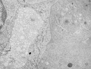
100 days after the invusion of artificial blood into mouse, this sample was taken of bone marrow. The white bubbly area on the lower left middle is typical of what perfluorochemical emulsions (at least those that eventually left the body) would look like inside phagolysosomes. The perfluorochemicals formed these small pockets in a sense re-emulsified within membrane bound structures in most cases (some perfluorochemicals like perfluoroiodide excepted). As a general rule of thumb, the faster the perfluorochemical blood went from large messy phagolysosome into a neat tidy highly ordered bubbly phagolysosome, the faster it left the body. I have been thinking on these images for 30 years, and hope to compare different chemicals in this respect. These are electron micrographic data from long past, before this type of blood substitute was approved and then disapproved for human use in the US. Nevertheless it tells us much about the immune system and processing of foreign materials (even man made materials).
 There are four types of instability in emulsions: flocculation, coalescence, creaming and Ostwald ripening…. all fun words that will likely apply to the transition of the first phagocytosed emulsion flocculant from the blood. I think in the case of the fluorocarbon emulsions there is a REVERSE Ostwald ripening in some, or alternatively, there is a minimum size that the lysosomal enzymes which are also within these phagolysosomal bodies can re-emulsify the fluorocarbon. (black dot is dirt… i didnt photoshop it out)