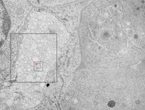
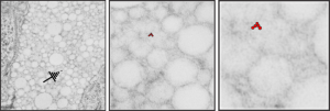
Electron micrograph of mouse bone marrow 100 days after the infusion of FC47 emulsion of perfluorochemical, in a study in the LC Clark, Jr, laboratory on artificial blood, blood substitutes. It has always been amazing that the original blood substitute-emulsion particles first are engulfed by phagocytes, and then demolished into seemingly amorphous inclusions, then with time, some perfluorocarbons, like FC47 are re-emulsified within the phagolysosomes into oodles (no clue how many millions) of small, very small and even smaller droplets. This particular electron micrograph of a bone marrow cell (just a portion of the cell) is packed with re-emulsified FC47 droplets within a membrane bound phagolysosome (for want of specifics on the enzymes in this organelle). It has been cropped and enlarged four times, and still there is no visible end of the increasingly tiny droplets. (Conjecture takes them to the level of the molecule. haha)
Arrow in top figure points to the small droplets that will be visible in the last of the enlargement inset figures.


Mouse: emulsion FC47 = perfluorotributylamine number 800325EI, IP, 50cc/x 7 =350 cc/kg (one whopping dose), I would have to look up the mixture, but typically 10% by volume with 5%F68, neg 5402, block 16203, 100 days post infusion, bone marrow, orig mag 13900, V, x 4: transmission electron micrograph. To estimate size, if a mammalian ribosome is between 25-30 nm in diameter on the same micrograph, then the size of the smallest red perfluorotributylamine droplet in the last inset is something around 10 nm in diameter.