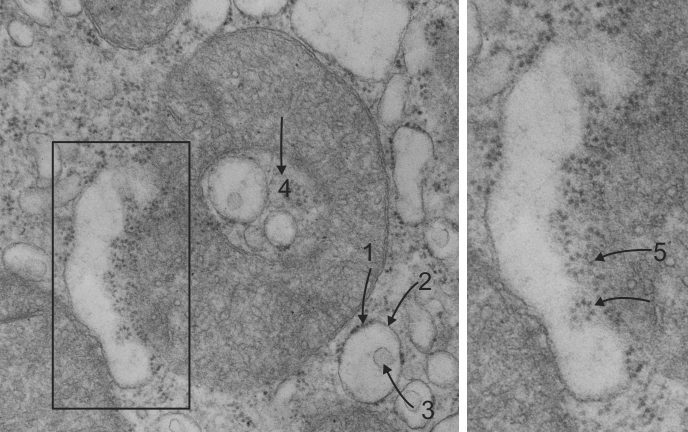
This is a great view depicting how closely aligned the outer mitochondrial membrane and the RER profiles can be. Smeared along a tangential section, opportunely showing about 7 ribosomes per spiral, (arrows 5) and also showing how close the RER is to the outer mitochondiral membrane and some of the cristi. So close, in fact, that it is not possible to define the separation between the two.
This sample is from mouse liver: indented (arrow 4) mitochondrion is indicative of oxidative stress, the RER components are dilated, some protein is seen, there are tiny vesicles within the RER profiles, ribosomes are not continuous (arrows 1 and 2), and have been lost at sites around each of the RER profiels. There are numerous polysomes, in addition to the ribosomal swirls attached to RER membranes. The increase in vesicles is a marker for the genetic disposition of this particular transgenic mouse. Mitochondria have a mix of tubular and flattened cristi, not that many intramitochondrial granules (Ca++ and Mg++). Right panel is an enlargement of black box in left panel.