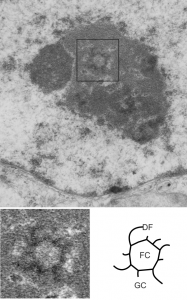
 The nucleus is a terrific place to do electron microscopy. Aside from the non-membrane bound elements within which increase the difficulty for any attempts at reconstruction in 3D (at least in my mind) the micro-environmental pressures push this dynamic organelle to rapidly change and adapt. The nucleus here had one particular area that caught my eye, that is the fibrillar center – dense fibrillar component (within the black box) of the top figure. The electron micrograph is from liver and it shows a fibrillar center with a circular (perhaps more octagonal) shape (as do most fibrillar centers) but the arms of the dense fibrillar component extend in a rotational pattern. In this case there are four long spiral arms and four short spiral arms (think “galaxy” with spiral arms). This is not seen often and it may be chance but not likely, in my opinion, particularly when one views the old time lapse videos of cells in culture and sees the nucleus and nucleolus spinning like little worlds in the medium called space. Rotational forces are alive and well within the cell and it is not a big stretch to witness their effects.
That said, here is a little fantasy (all else preceding is for real) — would galaxy arms of the dense fibrillar component in all cells go the same direction?, would it be clockwise, would the dense fibrillar component drag off to the counter counter-clockwise south of the equator, ha ha. For real, this is not a pattern I have seen described in the literature, nor recognized in the thousands of electron micrographs of my own, and thousands of others viewed, but I predict it will appear again and again.
Top figure is the original micrograph of a mouse hepatocyte, exposure evened in photoshop, one tiny scratch is patched (dare you to find it) without affecting interpretation. Black box surrounds the nucleolar fibrillar center and spiral arms of the dense fibrillar components which were copied and enlarged from the original micrograph. Inset lower left: burn tool in photoshop enhances direction of the galactic arms of the dense fibrillar component (which is visible in the original before emphasis), light central area is the fibrillar center. Diagram in lower right – stick figure for orientation, FC, fibrillar center of the inset to left, DF dense fibrillar component (galactic arms), and GC granular component. 12531_73229_#203 wc/ii liver 28d.