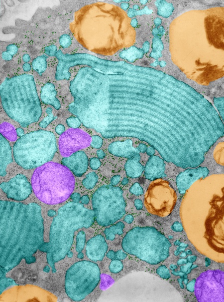
I am still trying to figure out what the ratio of ribosomes on the elongating end of these RER structures is. I think that the ribosomes appear to be staggered over the odd number of inner bands. Here is a jpg of a type II cell from a guinea pig (aged) which actually has a huge number of separate RER profiles stuffed with this protein (which I have pseudocolored cyan) and a few mitochondrial profiles (pseudocolored purple) a couple of lamellar bodies (kind of a gold color) and again, ribosomes here and there and plentiful at that, between and at the elongating ends of the profiles of RER.