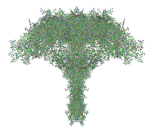
The more i look at the diagrams in yesterday’s post the more convinced I am that the surfactant protein A and D fuzzy balls are a molecules that evolved long before any nanostructure-production-designer molecules made by mankind aka the so called “globular glycofullerenes”.
Category Archives: Layered intracisternal protein granules in mammalian lung
Weibel-Palade bodies: fun stuff
I have seen these… at the present moment I am looking over the published literature for Weibel Palade bodies (WPD) in order to determine if there is ever a place where ribosomes are present on the bounding membrane. So far, none. This makes this cell adjesion group of molecules, while oligomerized into very beautiful structures, some like tubules, they are quite different from the intracellular granule purported to be a surfactant protein (possible SP-A) found in the cytoplasm of alveolar type II cells in the lungs of some species… which have a very precise localization and number of ribosomes on their limiting membrane (opposite sides of the long dimension of the granule, and approximately 4 ribosomes per 100nm).
| Weibel-Palade Bodies (WPB) | Layered granules in alveolar type II cells |
| At least 12 different proteins are found in granules | Undetermined number of proteins in type II cell granules |
| Recovered as oligomers in clathrin coated vesicles | Recovered, but not recycled or re-internalized as oligomers |
| Granules as single membrane bound SMOOTH ER | Granules as single membrane bound with end to end distribution of ribosomes, |
| Interacts with cell elements in blood | Interacts with fluid elements in alveolar space |
| Endothelial cell product | Epithelial cell product |
| Interacts with white cells and platelets | Interacts with alveolar macrophages |
| VWP apparently oligomerizes to influence the shame of WPBs | Oligomerization of the SPs involved in intracisternal bodes of alveolar type II cells create the granuel shape |
| WPBs maybe storage (albeit a small percent of total) for VWP | Granules in type II cells maybe for surfactant protein storage |
| WPBs have P-selectin in small quantitites, i wonder if these are for binding of clathrin proteins to initiate recycling from the secretion bubble structures after exocytosis of VWP? | Re-uptake, recycling of surfactant proteins is not so obvious (but happens) not big obvious vesicles… at least not that I have seen. |
Length, width, height, depth, thickness orientation?
This is hysterical. I wanted to describe the granule found in alveolar type II cells. I would have liked to use the distance between two outer dense layers of this regularly layered structure, which is 100nm as width, but find i need to use the word height. I had googled before which dimension is listed first, width or height for a standard rectangle…. and as i recall and as most people have given me measurements (yes for a different job, different hobby… making stained glass patterns) for width x height, and in that order… Portrait orientation being given as 8 x 10 or 8.5 x 11 etc etc.
So there is also the convention to use the word “length” for the longest a spect of a object. In the case of the protein granules in alveolar type II cells of some species, the long aspect can be many microns, or alternatively the granule can be short and stacked with many repeating “periods” having great height. If one uses only the “longest dimension” to connect with the concept of length then one gets into problems of orientation.
The question is then: what word do i use for the length in the direction parallel to the granule layering to describe it. And, do I use height or width to describe the 100nm thickness of the layered “period” or “periodicity” that can be a single unit or repeats over and over.
One would think that after thousands and thousands of years of language development that some very basic words for important stuff would have been established, but as i googled it, it becomes very apparent that they have not. For my purposes…. I will use the scheme above.
Other issues include convention by vocation, e.g., clothiers, tailors, lumbering, carpentry. In the graphics industry, it is width x height. As one blogger put it nicely (and to which I can certainly attest) best ask to be sure, otherwise it comes back to bite you.
Hexagonal pattern in protein granules in alveolar type II cells of the ferret
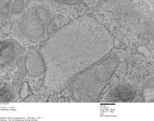
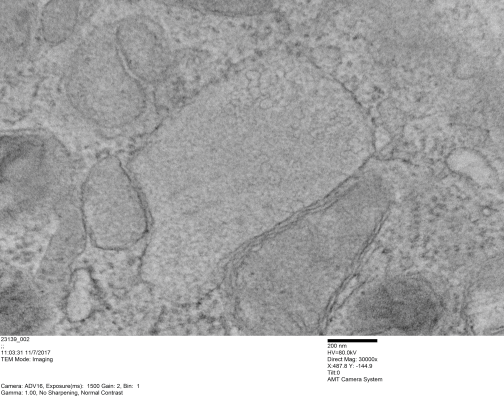
Hexagonal pattern in protein granules in alveolar type II cells of the ferret — yes still working on this. This image is from new sections, new microscope, but the results are the same. Image on top is unretouched, image in bottom i have burned what i see as a hexagonal pattern in this layered intracisternal granule.
Happy with this model of SP-A?
Here is a model of SP-A after weeks of trying to figure out which species (of the 7 looked at) to use, and where they are different and why the two programs I have used fail to predict the structure of the collagen segment… so i confess i am not a protein scientist, but seeing the really poor diagrams presented for this molecule (rather this octadecamer) by some very hefty scientists right here in this same University (no names mentioned here, though it would be my misguided pleasure to do so) I was determined to craft something that really might look like the real thing. Thus… the diagram below. The whole top part (mostly the carbohydrate recognition domain and coiled coil neck) was easily found as a pdb file and rotatable as a ribbon diagram too, and worked using the sequences for mouse, ferret, guinea pig and dog…. they are very similar, with a few pointy and craggy places along the top (presumably those places for recognition of organisms in the lung) and other immune functions. Coiled coil regions modeled particularly nicely, almost like rhythmic segments , and aptly called a neck…three or so bumps. These were also pretty similar among species. No problem there. But where the neck region meets the collagen-like stretch it just didn’t model the way (literally ALL) the diagrams represented it– except when i used it in the protein structure databases without the neck and CRD domains and part of the N terminal. Even then, most often that stretch of collagen-like sequence, even with the N terminal region added or subtracted, did not align with the suggested diagrams. Where, and how kinky the kink is IN THIS DIAGRAM is “speculation, and information from other diagrams” used here rather than the protein modeling (which didn’t model a “kink”).
So I pieced together a separate prediction for the collagen-like and N terminal sequences, and then “cut and pasted” the single protein into trimers and octadecamers, giving them the perspective that I was hoping to get by modeling the molecule as a “whole” and using the 3D options to rotate. Take this model as something synthesized from available models protein modeling software and available published diagrams.
The octadecamer is likely about 25-35nm across the top, and a similar height (which apparently changes with the spread of the CRD depending on open or closed conformation. These measurements are according to references found in the literature.
Top diagram, the perspective provided for the three angles that the trimers might be found in if they are as orderly (as indicated by the transmission electron micrographs i have posted. (Remember this is a partial-reconstruction and a partial diagram). Left view, kink facing forward, middle view, kink slightly rotated to side, right view is SP-A CRD top, neck slightly wider than the collagen and N terminal segments below.
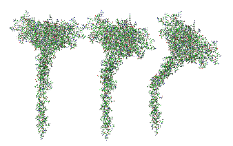 And here is the whole octadecamer, ordered with the three braided collagen-like domains aligned at the N terminal (as the literature suggests) then mirrored (from three above images) to make a total of 18 individual molecules of my generic SP-A).
Little pointy part of CRD in guinea pig SP-A
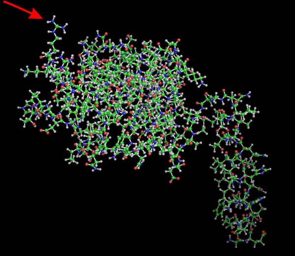
No laugh here, but I bet this little amino acid has a lot to do with some of the function of the carbohydrate recognition domain. Here is a Pymol view of guinea pig SP-A from residue 51 to 347, rotated to show this AA. Someone out there knows exactly which on it is… i will try to find out. In addition, I will post the ferret sequence from the same location (also residue 51 to end) to see if there is a similarity. RED arrow points to the spike.
Ball and stick model of surfactant protein A trimer
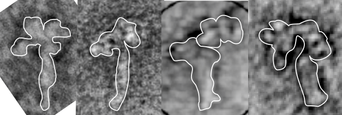
OK, apologetics first, i am not a protein chemist. But I ran this molecule in its entirety (it got truncated by the programs) so I diced it up into many many pieces, running each separately and trying to figure out how the pieces might fit together. This diagram (on the right) is the one I will use to create the octadecamer, the ball and stick diagram on the right is what i think is pretty accurate (and it matches some, but not all of the images I found in publications on SP-A, and it is just a little more accurate than those that make the CRD unit a round ball and the neck region a spring. It seems that only when I get the structure of the collagen-like portion along with the coiled coil of the neck to i get a structure that sort of fits what is seen with shadow cast TEM images (also published in peer-review journals). So this is what I am using. feel free to criticize. ha ha. 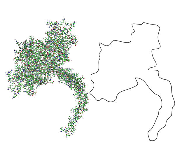
Here is a composite of several shadow cast images of SP-A, you can see that there is similarity. The dissimilarity with the ball and stick model might come from the fact that there are two configurations, open and closed, and that the closed configuration (which looks like the shadow cast molecules are in) is related to ion concentration. Almost all models of SP-A say there is an important “kink” in the collagen-like portion, and I am guessing that this is important for the images seen in traditional TEM of the alveolar type II cells which contain putative surfactant protein-A granules.
Pseudo-colored alveolar type II cell granules, and unretouched, and composite
Pseudo-colored alveolar type II cell granules, and unretouched, and composite image. I am just about finished with this study. I submitted the manuscript, I do hope if flies without too much revision. I am very aware that it is totally descriptive, but then descriptive science was the first science and it has great value. We all learn by what we observe, more so than what we propose (hypothesize) and test.
Here is a granule from my fav guinea pig #301 neg 9837, block 17084, which has several stacked parallel periods of the layered granule all in pretty nice array perpendicular to the layering. I extracted and then rotated the cropped portions, and increased contrast, and added half transparency to these four seemingly ideal areas. I merged them (with their transparencies matched as best I could) and flattened them as a fifth inset. This image is the inset in pink with 100 nm markers vertical and horizontal. The individual crops are shown below, colored just for fun.
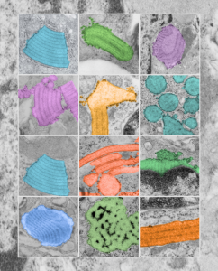
..yes, one more cover submission illustration — I just love these granules
..yes, one more cover submission illustration — I just love these granules, and I think this might be my favorite yet. Colored all the granules in (using photoshop) overlying their own original images from the basic research on this putative SP-A granule, and arranged them in a highly ordered format (haha) which is certainly not really my normal MO, since I am kind of a random thinker. But this is nice…in my opinion, and also a little artsie-craftsie for a scientist.. but thats what covers for scientific journals are for.
…and yet another cover submission for the manuscript on SP-A granule and electron microscopy
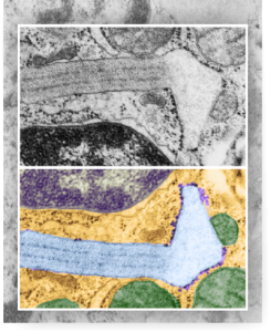
…and yet another cover submission for the manuscript on SP-A granule and electron microscopy. This one is a little more dramatic than yesterdays post from the bottom right part of the image. I like it better. It shows the quintessential granule, in black and white at the top, color at the bottom in a vertically mirrored image, directional ribosome attachment, electron-lucent areas under the non-oligomerized protein part, distinct periodicity to the dense bands (three complete “periods” in the lengthwise portion of this granule) and also the periodicity to the dense layers (3-4 dots/100nm) and even some of the faint periodicity found in the central lighter layers. In this electron micrograph one also sees part of the nucleus (bottom), a couple of nuclear pores, and some perinuclear space (but no granule formation in the perinuclear space is seen in this image, but it is found there frequently) and on the top, portions of three mitochondria and one adjacent to the lucent area of the granule. There are also other profiles of RER, coming and going which at some point might connect up to layered putative SP-A protein granule structures.
