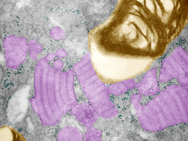
Working to determine whether there is an order to the ribosomes at the growing ends of the intracisternal proteins that are found in some aged animals of this and other species, I pseudocolored another transmission electron micrograph, intracisternal protein bodies are purple, and like the other images, the ribosomesa re cyan, and the lamellar bodies in the type II pneumocytes are gold-orange colored. It seems like there might be about two ribosomes on either side of the scalloped growin end, just before the darkest of the bands running perpendicular to the growing end. I can provide links to images that are very high resolution (26 megs) if you ask.