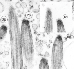
I just found these nice electron micrographs digging through the stack of 20k images and they are of the stomach of a mouse that is null for the gastric HKAtpase alpha. This was part of a study long time ago with many other main authors. The tips here are in the stomach, which was one of those unusual findings, that is, ciliated cells in and among the parietal and zymogen and mucus cells. Clearly ciliated however. So the tips of these cilia are really nicely preserved and in focus and it is clear that there is a glycocalyx, not exactly like that of the brush border in the intestine, more like that found in the trachea, typically “normal” ciliated epithelium. So below are TWO images (one inset) tips of 5 cilia which clearly show 6-17 filaments, and my projected total is given as well. The electron dense “sphere” or “dome” of attachment subadjacent to the plasmalemma is very obvious. 16447 and 16448 block 65165 gHKAa-/- mouse
I googled images to see what current research had been done on these fine filaments and the density seen within the tip of the cilia in the first figure below and found a reference back to 1978 (Charles KuhnIII, Wayne Engleman The structure of the tips of mammalian respiratory cilia Cell and Tissue Research January 1978, Volume 186, Issue 3, pp 491–498) which put the estimate of the number of filaments at something around 6 or 7, which makes no sense to me in 3dimensions…. So in the circles I approximated what i think the number of filaments would be. I found a second “antique” paper (only calling them antique because I have papers that old as well which contain details no t really found about ultrastructure in current literature) and it also thought the number of filaments was in the single digits. This diagram by Spicer (which of course I would redraw with more filaments). It is cute.
Sadly, a more current paper also quotes a low number of filaments… but has an interesting observation about a periodicity, so a quote from them is HERE “The apical structure of human respiratory cilia was studied using thin and thick sectioning techniques. The distal end of the mature cilium is narrowed and presents dense material and a crown-shaped glycocalyx. The crown is composed of three to seven bristles, having a mean length of 31 nm and showing a periodic structure. The capping structure of the apical part of human respiratory cilia is composed of a plate and an electron-dense cap tightly bound to the central and to each of the nine peripheral A microtubules. Lateral spokes link the peripheral microtubules to the internal ciliary plasma membrane and dense filaments radiate from the dense cap to the ciliary crown. These ciliary apical structures may be involved in several functions associated with microtubule assembly as well as ciliary movement and mucociliary transport. Apical structure of human respiratory cilia. Available from: https://www.researchgate.net/publication/19463484_Apical_structure_of_human_respiratory_cilia”
