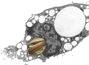
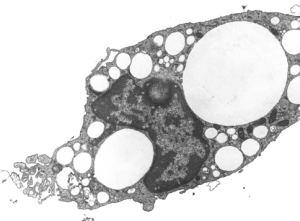
I really am fond of being silly, particularly when images like this hungry alveolar macrophage with eye and open mouth show up in my files. I probably took this image for my archives on liquid breathing way back in the late 1970s, never seeing the humor in the anatomy. But this morning when I was trying to figure out how to post this image, i just laughed out loud to see the perfect eye and open mouth and could not resist adding a “big mack” and “eyeball” with photoshop. The original, actual serious micrograph is below the cartoon.
So here is an alveolar macrohage, in a pretty good looking alveolus, from a mouse which was dunked in E2 for three hours (bubble oxygenated) and survived and was allowed to recover for 17 days. The pattern for E2 clearance from the lung is a little unique in that the droplets of perfluorocarbon are neatly coalescing into bigger and bigger droplets in some instances, but in others are seen as small droplets within, what i used to call “black cap lysosomes” which were apparently lysosomes with dense collections of enzymes, pushed over to one side of the organelle. Thus is was assumed the perfluorocarbon droplets became part of the lysosomal pathways. The E2 droplets here are white, I did not pseudocolor them as in the previous post, I did remove three pieces of junk using the stamp tool in photoshop. In this case the “eye” or “eyeball” seen for real in the bottom images is likely a tangential section of a medium size E2 droplet just pressing in on the nucleus making a symemtrical indentation. It does not have the character of a nucleolus, which the other option would be. The mouth of the cartoon was a reasonably large droplet of E2 (before i filled it in with big mack) probably on the order of 3-4 microns in the observed diameter. The tiniest droplets of E2 can be seen here and there in the cytoplasm, some of them even less than 0.5 microns in diameter. Mitochondria in these macrophages do not seem to be stressed by the presence of E2 droplets (more or less confirming their alleged inertness) and the amount of nuclear condensed chromatin would also suggest that the macrophages are not activated by E2 since there seems not to be any ramped up RNA, ribosomal, synthesis in the nucleus, or protein synthesis in the cytoplasm, which would suggest an increased synthetic activity related to an immune response to the presence of E2.
 And here is the scientifically sound, non cartoon, image.
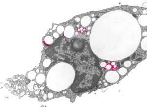
 Here is an image with four droplet or droplet groups which show the lysosomal enzymes highlighted in red. These observations relate to perfluorochemicals and fluorochemicals in general as the ability of the body to rid itself of them (whether through outgassing through the lungs reportedly in some relations to the vapor pressure and lipophilicity, and molecular weight. Other elimination is not reported to exist since perfluorochemicals are supposedly not metabolized, so metabolic degradation may not play a role here. I appears that how well the lysosomes “re-emulsify” the droplets is also relevant. Case in point for FC47… it really never was successfully re-emulsified, as opposed to perfluorodecalin which made nice progress in the process of forming tiny droplets in pretty uniform size during elimination. Here you can see the even roundness and lack of droplet substructure and marginalization of the lysosomal enzymes — all important characteristics of the lesser-evil perfluorochemicals.
