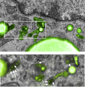
I posted a few days ago (here) a phrase which i thought was a little off color, and definitely made for effect, but stirred up something of interest in the way that perfluorochemicals “slippery and inert” might create or produce unusual lysosomal structures. I did post a composit lysosomal structure which looked to be like a little pouch full of varying size beads, or droplets, which are presumed to be the footprints of perfluorochemical left after liquid breathing or invusion of PFC emulsions. I examined some additional lysosomes this morning and it is clear there are many tiny droplets that are mixed in among some very large droplets, and these are so close to the nucleus, and near, perhaps almost looking like a connection, some of the ER in the cell which could be classified as RER. PFC ispossibly moving back up the ER profiles, and while all the names for endosomes (early, late, hybrid, budding, bla bla) this system is obviously continuous and naming “early” “late” etc adds little to the understanding (in my humble opinion), but naming them by pH and activities and/or membrane proteins associated, would be beneficial. Another issue is why there is a distinct dark band around the PFC droplets, it gives the appearance of TWO trilaminar membranes rather than a typical membrane bound endosome or lysosome.
Below is a micrograph (actually a scan of the original negative since the print I had marked and draw all over) which has two magnifications. Top image has white box which is enlarged below it, and the long stringy lysosomal structures are very close to the nucleus, even adjacent RER. this is almost like the “egg in snake” appearance. Presumptive perfluorochemical droplets (in this case E2 — structure given HERE) are colored green, the most electron dense areas surrounding the green (darker green) are lysosomal enzymes (also presumptive since I have not done any specific staining to verity it… but it is obvious.) White arrows point to the tiniest droplets (some in the range of about 30 nm, and some of those light areas may be tangential cuts off the ends of larger particles.
I suppose it is possible that the movement of PFC up the RER is to be expected because of its spreading chemical properties (at least according to Jean G. Riess and Marie Pierre Krafft.