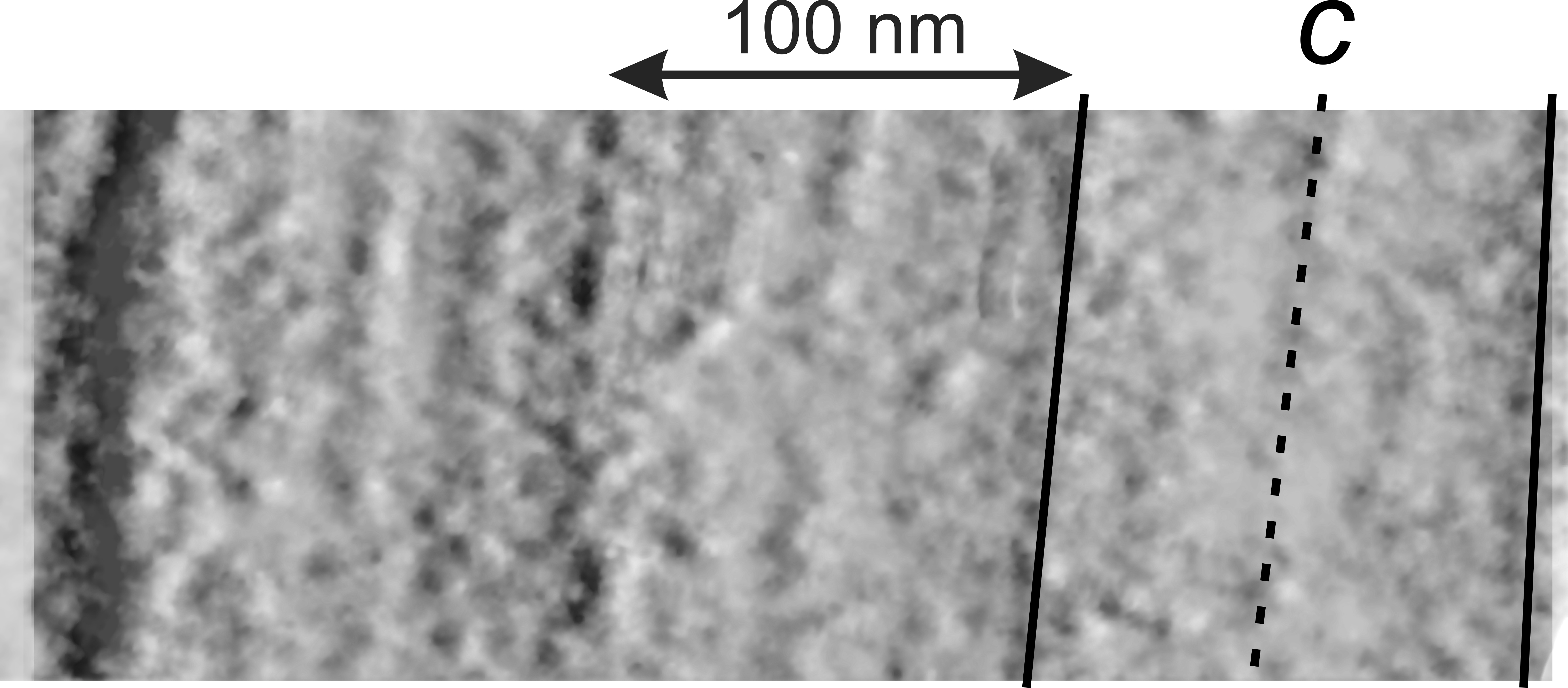
Here is a kind of interesting view of the bands (layering or periodicity) in the intracisternal bodies I am trying to figure out. This images is created by using a positive and negative image of the identical area, changing the transparency on both to about 80% then taking the negative image (lower layer) and moving it just laterally to create a shadow behind the positive image…. giving the whole thing a dimensional look. This clearly shows four distinct areas, with a central layer (band), and two identical bands on either side. This speaks perhaps to repeating but mirror arrangement of the molecules along the long axis of the RER membrane. Unfortunately I cannot remember if this is ferret or guinea pig.