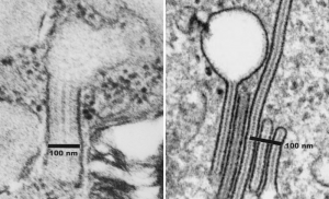
These two images, left is the same alveolar type II cell RER encased cisternal protein that has been made into a short edu-video on this site, and the one on the right is from Grassi et al, J Leuko Biology vol 64, 1998, which shows an intracisternal protein in a Langerhans cell (immune processing cell in the epidermis) called a Birbeck granule. Some similarities, but many differences exist between these two particular collectin-protein-layered-RER inclusions. The 100 nm marker shows the big difference in the size of the “between-RER-membrane boundaries for a single period, the one in the type II alveolar cell being more than 2 times wider. Here is an image, and also a new video.
