I have been trying to figure out why I am not able to find good electron micrographs which show all the interesting structural elements of mitochondria. Not just the trilaminar membranes that make up the inner and outer membrane portions, and the matrix and the intermembrane space and the granules, those are easy to find. What has been a real hunt is to find mitochondria where there is some element that could be called a mitoribosome (supposedly situated on the inner mitochondrial membrane), or a portion of circular DNA (supposedly situated in the mitochondrial matrix). These are structures in the nm size range and should be able to be seen. Some mitochondrial respiratory chain molecules can be seen – HERE and HERE which might be ATP synthase, but not the complexes I – IV and others. So apparently not much in the matrix such as described as poly-mitoribosomes, or proteins being synthesized (as sometimes can be seen in the cytoplasmic RER gets appropriately labeled. Diagrams are particularly disappointing as well.
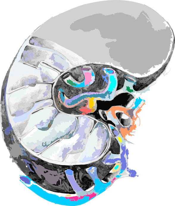
Here is a drawing from years ago, I am going to work to add substructural units in relative size and see if that helps. cut-away of a mitochondrion, with tubular ER on the right.

HERE is an article that answers some of my questions.