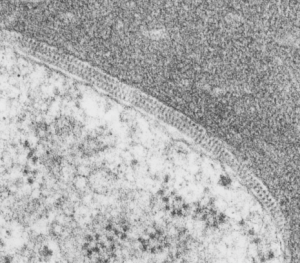
Intracristae – membrane protein organized in mitochondria — found in guinea pig liver electron micrographs in a retrospective study on vinyl chloride (and air) and vitamin C exposure from the 1980s. These cristae with apparent organizations of ATP synthase in a long row, parallel and sometimes looking staggered, sometimes aligned, have appeared in exposed as well as control (both groups receiving vitamin C). This is an awesome image with a 3-layered row of highly organized molecules looking a little like they are strung together somehow, yet each distinct. Just marvel at the order in the completely UNRETOUCHED electron micrograph of a mitochondrion in guinea pig liver here.
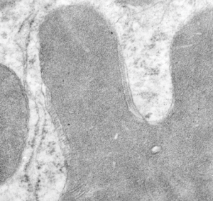
 Mitochondria in both control and experimental data sets have mitochondria with wild shapes, very atypical for mitochondria in general, in some cases on section looking like asterisks (yep, same type outer membrane configuration seen in apoptotic cells). This is the case of the mitochondrion shown enlarged (the scalloped edges are just cropped so the ATP synthase is more visible. Image below 5924_17010_guinea_pig_air_vitC_liver.
As with the intracisternal body in the alveolar type II cell of the lung of many guinea pigs, when the protein within the cisternae of the RER begins to assume a substructure (in that case, a layered granule), then the resulting RER becomes very rigid looking. Similarly here, when the cristae have the ATP synthase all aligned in alternating double rows, the intramitochondrial membrane and other elements in the mitochondria also take on a rigid appearance.
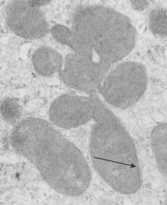
 Micrograph below shows a section of an example of odd shaped mitochondrion with an area of extended ATP synthase molecules head to head, and on the inner mitochondrial membrane a little bit of rigidity and a thickening of the membrane is seen (arrow).
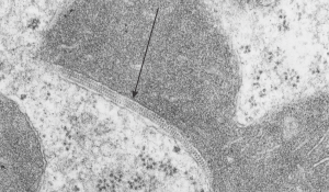
 Here is a mitochondrion with an extended section of rigidity in the mitochondria membrane adjacent to a TRIPLE row of ATP synthase molecules, lucky cut (I hope that is what this is), and again rigidity in the mitochondrial membranes in this zone, and inner mitochondrial membrane has a slightly increased electron density about it (which of course means something, but I don’t know what).

HERE is a nice article…. it says the protein is in the matrix, which is not what is seen in the micrographs above, it is in cristae…. so there is some explaining to do on this topic.