This looks simple, but it took a while to figure out… but I can use CorelDRAW for all measurements. This program is amazing and I could hardly survive without it. Thanks to my son danny for introducing me to it way back when he was just a kid working at a deli that for some reason used one of the very first versions… CorelDRAW 3. I should buy the upgrade (haha). So measurements of molecules and structures that have been photographed with electron microscopy (or atomic force microscopy, or techniques which shadow or coat molecules to highlight them are rarely made. Some of the really great “oldies” that formulated the basics for morphometry inspired me 40 years ago, and making what is usually a “subjective and only visual” science into something that can be graphed and analyzed has been my goal (forever it seems). So working on the shapes of two surfactant protein molecules (SP-A and SP-D) it became clear that no one really paid much attention to how the represented these molecules graphically… some even represented (and published) them with such “license” (which certainly could NEVER be considered “artistic license” so erroneously that it was adding misinformation to other articles whose authors were using them as models. FAKE NEWS.
It has been great fun working with images of surfactant protein D for TWO reasons: 1) the totally ridiculous diagrams that have been published… i wanted to set straight with something more accurate and 2) these molecules can be very important in the efforts to tailor new synthetic – fullerene-type innate immune molecules to best opsonize viruses and bacteria which enter the lung.
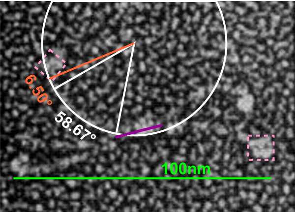
Previous posts show center arc angles in dodecamers, and also the angles of the collagen-like portion of the SP-D dodecamer “arms”, but other things need to be quantified. Diagram below indicates what I think i can measure, and that is the arm length of the collagen-like portion, maybe also the length of the coiled coil neck region, the varying shape-size of the CRD, and the mean length of the N terminal. PER THE IMAGE below.
Using the center arc angle of the collagen-like portion the formula found on this site can be used to determine the arc length. Similarly, the neck arc length. the purple line is from the branch of the dodecamer pairs, and it seems actually to vary quite a bit. THe CRD are easy to measure just as with width and height (since it is a trimeric structure (anticipating from the molecular models on RCSB to NOT have equal width and height, but vary in orientation sufficiently in the micrographs to present with some variation.
There are two kinks for sure, one was predicted by some researchers to be close to the N terminal, and I see one (sometimes represented in various diagrams) at the coiled-coil neck region. I havn’t decided whether to measure that or not. So many studies have shown that the dodecamer is about 100nm that has been used as the standard measure from which other measures are made. EDIT: so neck measures are not going to be possible, and will be part of the CRD measurements.
