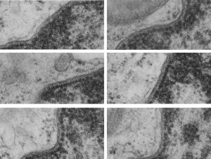
Just looking at a single nucleus, single inner nuclear membrane (INM), single cell, it becomes quite clear that the organization of the INM varies and that three very prominent differences in that organization are apparent. A normal configuration (I am only calling it that because it is “in between”, and a very dense organization, and a loose and spacey organization (see previous post on june 24 2016). The image below is something of a quick and easy demonstration of that point. Electron micrograph of the inner nuclear membrane was cut in six different places from an image of a dog type II alveolar pneumocyte, a control animal from experiments decades ago, and It was easy to see three types of organization. tight, middle and loose. It will be interesting to determine which processes go along with which anatomical arrangements (though the outcome is not difficult to predict).
Top=two segments of the inner nuclear membrane which is “neither tight nor loose”, and orderly; middle row=two examples of “tight” organization and bottom row, two examples of “loose” organization. Inner nuclear membranes all coursing upper right to lower left, condensed chromatin is on the right hand side of the nuclear membrane, none of the profiles contains a nuclear pore, the slightly less dense layer of lamin is present in all images, some areas of euchromatin are seen, upper right image has a portion of a lamellar body, two other images each have a portion of a mitochondrion, scattered ribosomes are on the left hand side of each image, and the perinuclear space is not particularly dilated in any of the images.
