Previous comments about some kind of order – periodicity – linearity? in the micrographs of hepatocytes which contain inclusions of perfluorodecyliodide (IPD ) and there are others that I noted. (nb. quote)Ubiquitous, highly glycosylated, integral membrane proteins of largely unknown function, called lysosome-associated membrane proteins (LAMPS) or lysosomal integral membrane proteins (LIMPS), account for about 50% of the protein in the lysosomal membrane.
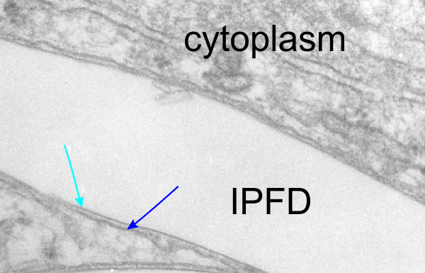
1. the trilaminar membrane has an exaggerated density on the outer membrane, and a very smooth (maybe no integral or surface proteins) on the lamina of the membrane in contact with the IPFD. See picture below, blue arrow inner trilaminar membrane (maybe few proteins) outer trilaminar membrane (red arrow) with maybe lots of membrane proteins. It would be interesting to nkow for sure if the IPFD causes this lack.

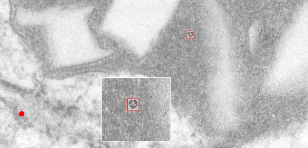
Another interesting thing is that there is a substructure to the lysosomal protein matrix — in fact at least three different organizations that I can detect, and this one just happens to match up in terms of size with LAMP-1…. for which there are shadowed images by Carlsson and Fukuda of the dimer found here which i have sized using their micron bar marker and the size of a ribosome (used 25nm) and superimposed over a giant lysosome found in the liver of a mouse that had been infused with IPFD (perfluorodecyl iodide). (see below) Tiny red box (enlarged in inset) is their image of the rotary shadowed dimer of LAMP-1, which i have placed into the textured density of one portion of a large lysosome containing IPFD crystals. Red dot is an aproximate size of an hepatocyte ribosome (25nm used). Wouldn’t that be a fun thing to spend some time figuring out, since I have only suggested it, not verified it.
