Here is an interesting publication which I attempted to use to demonstrate an easy morphometric method for defining AFM images.
This is apparently an amaloid protein which the authors called apo-SBB fiber and I personally know nothing about this protein but have made some measurements that show that interesting things can be derived from using simple morphometric tools to evaluate microscopic images.
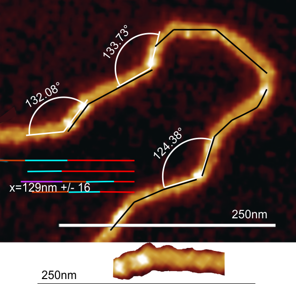
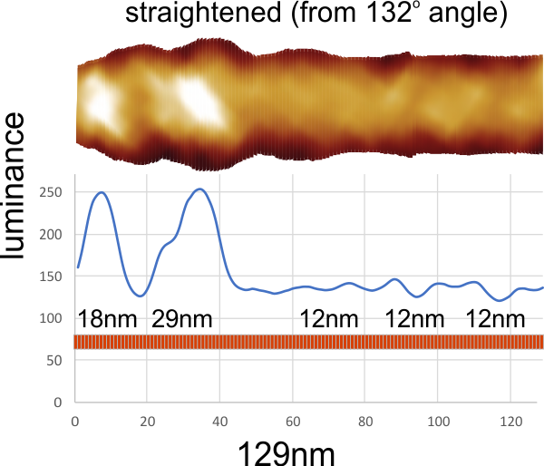
It appears that the peak heights (luminance) might be about 12-15nm in “diameter” obviously not round here. In the images measured below there is not number for the bar micron marker provided, but it has been estimated from the “diameter” measure above to approximate a reasonable number — which looks to be 50nm.
The peaks do not appear to me to be related to the bends (the 129o angles). Some peaks are adjacent to bends, some prior to bends, and others right at the bend, thus in these few images provided there may not be a predictable pattern to the peaks except that about half of the peaks appear as two nearby peaks, the other half are singlets .
Measuring arc angles from 13 angles on the above and below images stayed similar to what was obtained for the single colored micrograph above, it change only slightly to arc angle=128.3 decrees +/- 10
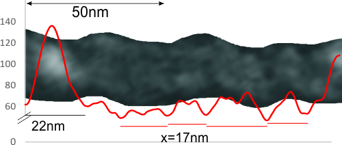
It also seems like there is a periodicity in the protein length as well.This portion of a fiber is taken from the most faint of their tracings which to me showed amazing twisting. The periodicity (shown above at about 12nm and here X of 17nm probably somewhere between, and the peak shown above two different measures, and here at about 22nm which is close to above (despite their lack of marking their bars with actual micron numbers). So the different mags, and measures look similar for their pictures. I did NOT need to straighten this particular portion, so that also confirms that using the slice and center technique does pretty well ad straightening out images of proteins that are curved.


