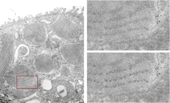
Ferret alveolar type II cell RER inclusions (layered protein granules) appear as evenly stacked bodies, or often on tangential section, areas smeared out concentrically or linearly. These intracisternal bodies, which i think I should begin to call surfactant protein granules (ha ha, not to rile up any feathers) have many subtextures and periodicities which show up when I examine my stash of electron micrographs close up. Bless my scanner and photoshop, as they allow me to crop and highlight and darken and make what I see initially, into something that can be highlighted for everyone to see. Such is the case with several of these intracisternal protein surfactant granule bodies found in a ferret alveolar type II cell. The original cell is shown in the video, and a small inset is enlarged, and the periodicity of the protein (which I think is an accumulation of surfactant protein A — maybe upregulated in production as an immune response? I don’t know) along the dense bands of the 100 nm periodicity. The lighter bands don’t really show up very well on tangential sections, though I have some micrographs where some substructure is visible. The images used to make the video below is here as a png file, where the enhancement of the periodicity can be seen in the figure in the lower right, the area from which this inset is taken is shown in the red box on the left. The entire ferret alveolar type II cell is on the left, apical border facing top. There are at least 6 inctracisternal body surfactant A granules? present, some showing parallel substructure other showing curved and tangential cuts.
Here is a short animation using these transmission electron micrographs