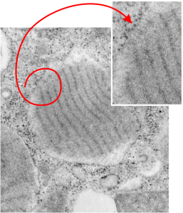
Electron lucent areas just beneath the RER limiting membrane alveolar type II cell, these have ribosomes and layered protein, just like the previous post. I suspect it has to do with the proximity of ribosomes to the protein beneath, and the lucent areas just seem not to have close-by ribosomes. The red circle highlights which part of the intracisternal body is being noted, and the arrow head points to the insert and enlargement of that arrow. As before, the original large image is unretouched while the inset has been “burned” in the area that I want you to see, which includes the more electron dense area and adjacent more lucent areas, and the ribosomes. This is an alveolar type Ii cell from a ferret, magnification is 19,500, enlargement before scanning is 4 x. Final magnification can be more or less approximated by the size of the ribosomes (reportedly 20-30 nm) which makes the dark bands within the intracisternal body separated by a distance of about 100 nm.
