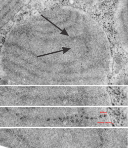
More periodicity seen in RER granules in alveolar type II cells from a ferret. In the areas of this single intracisternal RER protein there are great examples of the periodicity of the protein as it was fixed (this fixation was standard paraformaldehyde glutaraldehyde fluid). This particular electron micrograph is a little grainy (I probably used dektol in stead of microdol to develope it — in hindsight not a good shortcut to take, but sometimes we do things without knowing the 30 year consequences.. duh. At any rate, I have compared areas in the dense band with the lucent bands of this particular tangential cut through a curved RER granule also called intracisternal body and shown the unretouched (in terms of the densities) and the burned densities as I could see them. There is a portion where there is not periodicity taken from the exact same RER granule less than 100 nm below the string of beads seen above it. Ribosome (20-30 nm? is used to compare sizes, and the beads look to be something on the order of 50 nm.
Full granule is pictured in the transmission electron micrograph on top, arrows point to the horizontal segment which has the unretouched periodicity, the arrow below points to the lucent zone used to compare whether I am hallucinating the periodicity (ha ha). Micrograph 2, just beneath the full granules is enlarged unretouched (i did remove some dust and a scratch using the stamp tool in photoshop but in the line of granules). Micrograph 3, is a view of the exact same strip of the intracisternal body as image 2, but i enhanced the beads with the burn tool. and micrograph 4 is the more or less no-granule-zone in the lucent area as spread out in a tangential cut, unretouched.
