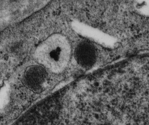
Original, and unretouched or processed (not even contrast enhanced) photo of a small profile of rough endoplasmic reticulum from the alveolar type II cell. It has two ribosomes over to the left about 10 o’clock, they can give you a feeling for the dimensions of the fuzzy balls within the central portion of the profile. The electron lucent area surrounding the dense center is pretty common in these intracisternal bodies (granules if you prefer) and the layering here is too large to be part of the 100 nm pattern seen when the bodies are found perpendicular to the periodicity. I looked closely at the center, and I would estimate that the bodies (in groups of three – yes, my guess – roughly) is something around 60-70 nm, at least bigger than the ribosomes (when lumped together in threes or whatever the cluster contains. OF course the whole granule is much bigger, but it would be fun if the smaller portions of the central density of this intracisternal body equated to 50 – 60 nm groups of three bouquets of surfactant protein A. That would be just fun.
You can find a short animation with text at this link to YouTUBE:
