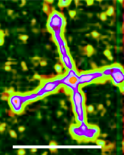
Doesnt seem to matter much what processing happens to these images. The data remain abundantly clear for SP-D dodecamers. N termini, glycosylation peaks and three small peaks in the collagen like domain and nice CRD. At this point: it doesn’t matter the direction, or the lines in the AFM, the image processing, or the angles between the trimers — it looks like three small peaks in the collagen like domain. Over and over again they show up. See tone curve graph in bottom image. SP-D imagesaved and processed from Arroyo et al.